Adrenal Disorders for NEET PG — Complete Guide 2026
Master adrenal disorders for NEET PG 2026: zona architecture, Cushing syndrome, primary aldosteronism, Addison disease, congenital adrenal hyperplasia, pheochromocytoma, and adrenal incidentaloma with diagnostic workflows.
NEETPGAI EditorialPublished 11 Feb 202621 min read
Share this article
This content is for educational purposes for NEET PG exam preparation. It is not a substitute for professional medical advice, diagnosis, or treatment. Clinical information has been reviewed by qualified medical professionals.
Ready to put this into practice?
Start practicing NEET PG MCQs with AI-powered explanations.
Primary aldosteronism — ARR >20–30 screens; confirmatory saline load / fludrocortisone test; AVS before surgery for unilateral adenoma
Addison disease — autoimmune (West) or TB (India); short Synacthen test peak <18 mcg/dL; high ACTH, high renin, low aldosterone
Adrenal crisis — shock + hyponatraemia + hyperkalaemia; IV hydrocortisone 100 mg BEFORE cortisol results, then 200 mg/24 h; IV saline + dextrose
21-hydroxylase deficiency — ~95% of CAH; ↑ 17-hydroxyprogesterone; classic salt-wasting (1–3 wk of life), simple virilising, non-classical forms
Pheochromocytoma — rule of 10s; plasma free metanephrines (high sensitivity); alpha-blockade first (phenoxybenzamine), then beta
1 mg overnight dex test — cortisol >1.8 mcg/dL next morning = abnormal (Cushing positive screen)
Adrenal incidentaloma — screen every mass for Cushing + pheo; add ARR if hypertensive; resect if >4 cm, functional, or imaging-suspicious
CT Hounsfield Units — <10 HU on unenhanced CT = lipid-rich benign adenoma; >20 HU = indeterminate → MRI chemical shift / washout
Adrenal disorder vignettes are NEET PG staples — the hypertensive hypokalaemic patient on no diuretic, the cachectic Indian man with hyperpigmentation and hypotension, the 46,XX neonate with ambiguous genitalia and salt-wasting crisis, the young hypertensive with paroxysmal headaches and palpitations. This guide covers zona-specific physiology, Cushing syndrome, primary aldosteronism, Addison disease, CAH, pheochromocytoma, and the adrenal-incidentaloma workflow. Pair with the medicine subject hub, the pituitary disorders guide, and the common mistakes in medicine NEET PG article for endocrine integration.
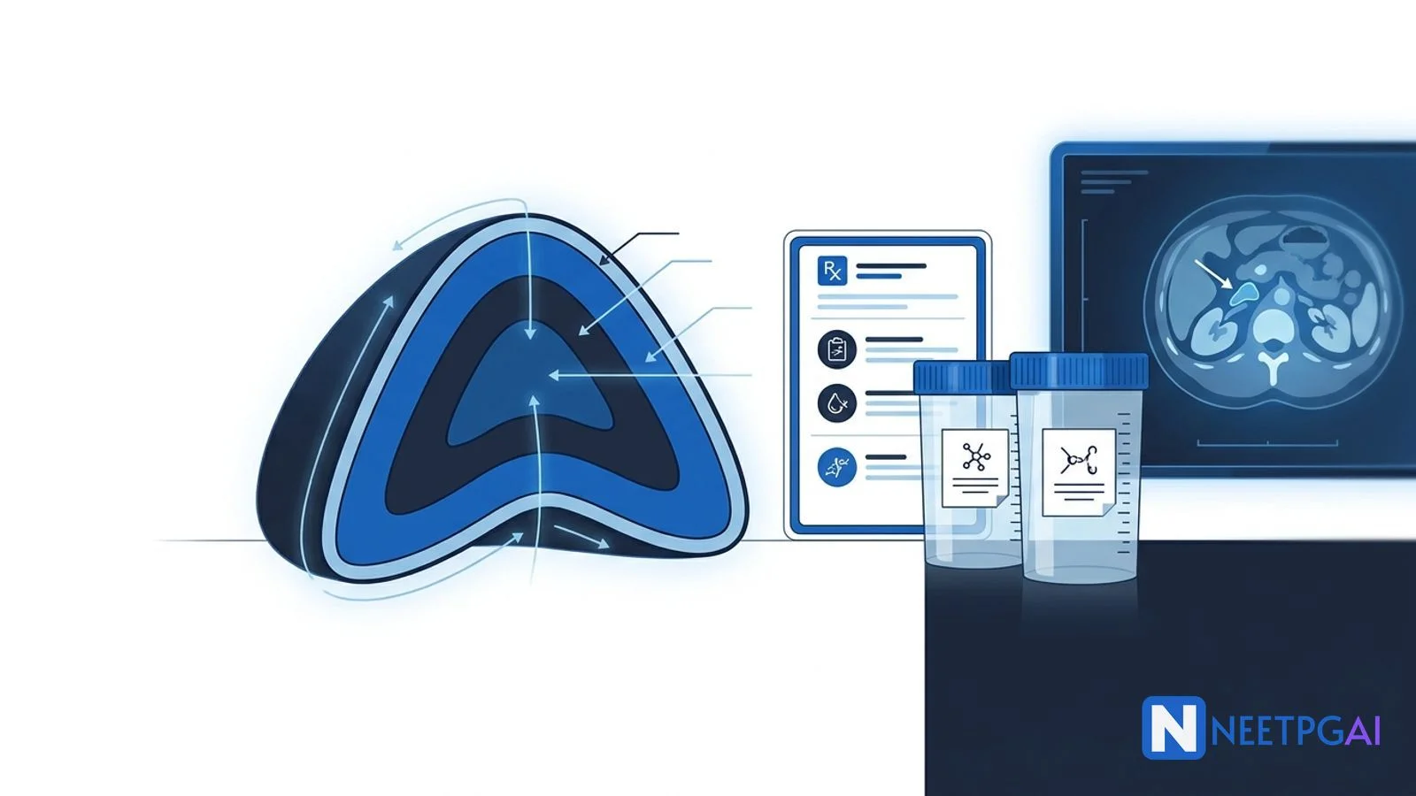
Adrenal anatomy and zona architecture
The adrenal gland is a bilateral retroperitoneal organ lying on the superior pole of each kidney, with an outer cortex and inner medulla that have different embryological origins and secretions.
Anatomy essentials:
Cortex — mesodermal origin; three concentric zones, each with a distinct enzyme repertoire and product
Blood supply — superior (from inferior phrenic), middle (from aorta), and inferior (from renal) suprarenal arteries; drainage differs on the two sides — right adrenal vein drains directly to IVC, left drains to left renal vein (surgical relevance)
Three cortical zones (GFR mnemonic — salt, sugar, sex):
Cholesterol → pregnenolone (rate-limiting; StAR transports cholesterol into mitochondria, CYP11A1 / desmolase)
Pregnenolone branches into three pathways — 17-OH-pregnenolone / progesterone (to cortisol and androgens) and progesterone (to aldosterone and cortisol)
21-hydroxylase (CYP21A2) converts 17-OH-progesterone → 11-deoxycortisol and progesterone → deoxycorticosterone (DOC) — its block defines the commonest CAH
11-beta-hydroxylase (CYP11B1) deficiency causes hypertension from DOC excess
17-alpha-hydroxylase (CYP17A1) deficiency causes hypertension and sexual infantilism
Medulla:
Chromaffin cells synthesise noradrenaline (NA) and adrenaline (A) — PNMT (phenylethanolamine N-methyltransferase) converts NA → A and requires cortisol from the portal cortical–medullary blood supply
Metanephrine (from adrenaline) and normetanephrine (from noradrenaline) are the stable metabolites used in pheo diagnosis
Extra-adrenal chromaffin tissue along sympathetic chain gives paragangliomas
Cortisol physiology:
Diurnal rhythm — peak 6–8 am, nadir around midnight
~90% protein-bound (CBG, albumin); free cortisol is the active fraction
Feedback — cortisol suppresses hypothalamic CRH and pituitary ACTH
Cushing syndrome is chronic hypercortisolism from any cause — exogenous glucocorticoids are the commonest cause overall; endogenous causes split into ACTH-dependent and ACTH-independent.
5–20 pg/mL — repeat and clinical correlate; consider CRH stimulation
Step 3 — localise (ACTH-dependent):
High-dose dexamethasone suppression (8 mg) — Cushing disease usually suppresses >50%, ectopic often does not (not absolute)
MRI pituitary — adenoma detected in ~60–70%
IPSS (inferior petrosal sinus sampling) — gold standard when MRI negative/discordant; central:peripheral ACTH >2 basal or >3 post-CRH confirms pituitary source
CT chest/abdomen ± 68Ga-DOTATATE PET for ectopic source
Management:
Cushing disease — transsphenoidal surgery first-line (remission 70–90% microadenoma)
Adrenal adenoma — laparoscopic adrenalectomy
Adrenal carcinoma — open adrenalectomy + mitotane adjuvant
Ectopic ACTH — resect source; if not found/metastatic, medical therapy
Medical therapy (bridge or refractory) — ketoconazole, metyrapone, mitotane, osilodrostat (cortisol synthesis inhibitors); pasireotide (pituitary-directed); mifepristone (GR antagonist for steroid-induced diabetes)
Bilateral adrenalectomy — refractory pituitary Cushing; risk of Nelson syndrome
Peri-op and post-op stress dosing; taper steroids slowly after cure (HPA axis recovery takes months)
Primary aldosteronism (Conn syndrome)
Primary aldosteronism is autonomous aldosterone excess — the most common reversible secondary cause of hypertension — and normokalaemia is the rule, not the exception.
Subtype (laterality) — CT adrenals ± adrenal venous sampling (AVS) — AVS is the gold standard before considering adrenalectomy (imaging alone misclassifies in up to 30%)
Age <35 with clear unilateral adenoma on CT + florid biochemistry — may proceed to surgery without AVS (AVS still preferred)
Management:
Unilateral (APA / UAH) → laparoscopic adrenalectomy (cures HTN in ~50%, improves in most)
Bilateral (BAH) → spironolactone (first-line; gynaecomastia side effect) or eplerenone (more selective, fewer side effects, but expensive)
Add amiloride, thiazide, salt restriction as adjunct
FH-I → low-dose dexamethasone
Addison disease and adrenal crisis
Addison disease is primary adrenal insufficiency — destruction of the adrenal cortex causing glucocorticoid, mineralocorticoid, and androgen deficiency — classically autoimmune in the West and tuberculous in India.
Causes of primary adrenal insufficiency:
Category
Causes
Autoimmune adrenalitis
Commonest in developed countries; anti-21-hydroxylase antibodies; polyglandular syndromes (APS-1, APS-2)
Infectious
TB (commonest in India), HIV opportunistic (CMV, MAC, histoplasma), disseminated fungal
Monitor electrolytes every 4–6 h (watch for hyperkalaemia correcting, sodium overshoot)
Fludrocortisone not needed acutely (high-dose hydrocortisone covers mineralocorticoid effect)
Transition to oral hydrocortisone once stable; restart fludrocortisone when hydrocortisone <50 mg/day
Congenital adrenal hyperplasia
Congenital adrenal hyperplasia (CAH) is a group of autosomal recessive disorders of cortisol biosynthesis — reduced cortisol causes ACTH-driven hyperplasia and shunting of precursors into androgen or mineralocorticoid pathways.
Enzyme blocks and phenotypes:
Enzyme (gene)
% of CAH
Cortisol
Aldosterone
Androgens
BP
K+
Classic feature
21-hydroxylase (CYP21A2)
~95%
Low
Low (salt-wasting)
High
Low
High
Virilised 46,XX / salt crisis
11-beta-hydroxylase (CYP11B1)
~5%
Low
Low aldosterone but high DOC (salt-retaining, HTN)
High
High
Low
Virilisation + HTN
17-alpha-hydroxylase (CYP17A1)
<1%
Low
High DOC (HTN)
Low
High
Low
Sexual infantilism + HTN
3-beta-HSD
Rare
Low
Low
Weak (DHEA ↑)
Variable
Variable
Ambiguous in both sexes
StAR (lipoid CAH)
Rare
Very low
Low
Low
Low
High
Severe salt-wasting, all steroids low
21-hydroxylase deficiency — three forms:
Form
Onset
Enzyme activity
Features
Classic salt-wasting
Neonate (1–3 wk)
<1%
Vomiting, shock, hyponatraemia, hyperkalaemia; ambiguous genitalia in 46,XX; 46,XY normal male genitals but adrenal crisis
Simple virilising
Neonate / childhood
~1–2%
Virilisation without salt-wasting; ambiguous genitalia in 46,XX; precocious pseudopuberty in males
Non-classical
Adolescence / adulthood
20–50%
Premature pubarche, hirsutism, acne, PCOS-like in women; often missed
Diagnosis:
Elevated 17-hydroxyprogesterone (17-OHP) baseline and after ACTH stimulation (>1000 ng/dL at 60 min post-cosyntropin)
Newborn screen (heel-prick 17-OHP) — practised in many countries
Elevated DHEA and androstenedione
Hyponatraemia + hyperkalaemia in salt-wasting form
Genotyping confirms
Karyotype + pelvic USG for ambiguous genitalia
Management:
Hydrocortisone 10–15 mg/m²/day divided TDS (preferred over long-acting in children to minimise growth impact)
Fludrocortisone 0.05–0.2 mg/day + salt supplementation in salt-wasting form
Stress dosing — triple oral dose / IV hydrocortisone for illness
Surgical correction of virilised genitalia (timing individualised, ethical considerations)
Prenatal dexamethasone to mother at risk — historically used to prevent virilisation in 46,XX fetus; now controversial (risks outweigh benefits for most)
Monitor growth, bone age, androgen levels, 17-OHP, PRA
Pheochromocytoma and paraganglioma
Pheochromocytoma is a catecholamine-secreting tumour of adrenal chromaffin cells — paraganglioma is the extra-adrenal counterpart — classic NEET PG vignette is paroxysmal hypertension + headache + palpitations + sweating.
Rule of 10s (classic teaching — modern data put familial higher):
10% bilateral
10% extra-adrenal (paraganglioma)
10% malignant (defined by metastasis, not histology)
10% familial (now known to be 30–40%)
10% in children
10% not hypertensive (normotensive pheo)
10% in normotensive screening series
Hereditary syndromes:
Syndrome
Gene
Associated features
MEN 2A / 2B
RET
Medullary thyroid ca, primary hyperparathyroidism (2A), mucosal neuromas/marfanoid (2B)
Localisation — CT abdomen with contrast (initial); MRI if CT negative; 123I-MIBG scintigraphy for metastatic/extra-adrenal/malignant; 68Ga-DOTATATE PET sensitive for SDH-related paraganglioma
Genetic testing in all patients (especially young, bilateral, paraganglioma, familial, metastatic)
Management — peri-operative:
Alpha-blockade first — phenoxybenzamine 10 mg BD, titrated up over 10–14 days (non-competitive irreversible alpha); or doxazosin/prazosin (competitive)
High-salt diet (5 g/day) + IV fluid from day 2–3 of alpha-blockade to re-expand volume
Beta-blockade — added only after adequate alpha-blockade if tachycardia persists (atenolol, propranolol)
Calcium-channel blocker — adjunct (nicardipine)
Metyrosine (tyrosine hydroxylase inhibitor) — occasional adjunct for large / metastatic disease
Surgery — laparoscopic adrenalectomy (or open for large/malignant); avoid tumour manipulation until vessels ligated; intra-op hypertension → IV phentolamine / nitroprusside; post-op hypotension → fluids + noradrenaline; post-op hypoglycaemia from rebound insulin release
Follow-up — annual plasma metanephrines; genetic counselling for family
Why alpha-before-beta: beta-blockade alone leaves unopposed alpha-stimulation (catecholamines now only bind alpha receptors) → paradoxical hypertension, heart failure, pulmonary oedema.
Malignancy — no reliable histological marker; defined by metastasis (most commonly bone, liver, lungs, lymph nodes). Managed with surgery, 131I-MIBG therapy, chemotherapy (CVD), targeted agents (sunitinib).
Adrenal incidentaloma
Adrenal incidentaloma is an adrenal mass >1 cm found on imaging done for an unrelated reason — prevalence rises with age (up to 7–10% in those over 70). Evaluate along two axes: function and malignancy.
Functional work-up (every incidentaloma):
Test
Purpose
1 mg overnight dexamethasone suppression test
Screen for subclinical / autonomous cortisol secretion (present in ~5–20%)
Plasma free metanephrines or 24-h urinary fractionated metanephrines
Screen for pheochromocytoma
Aldosterone-to-renin ratio
Only if hypertensive or hypokalaemic — screen for primary aldosteronism
DHEA-S, testosterone, 17-OHP
Only if virilising features or suspected ACC
Oestradiol
Only if feminising features (rare ACC)
Malignancy / phenotype assessment:
Imaging clue
Interpretation
Unenhanced CT <10 HU
Lipid-rich benign adenoma (very high specificity); no further imaging
Unenhanced CT >20 HU
Indeterminate — do CT washout or MRI chemical shift
Absolute washout >60% or relative washout >40%
Benign adenoma
MRI chemical shift (drop in signal on out-of-phase)
Lipid-rich adenoma
Heterogeneous, necrotic, irregular margin, calcification, >6 cm
Adrenocortical carcinoma suspected
Bilateral, history of primary elsewhere
Metastasis
Hyperintense on T2, high enhancement
Pheochromocytoma (always screen biochemically before any biopsy)
Size thresholds:
<4 cm — observe with serial imaging (6–12 months, 2 years) if functional screen negative and imaging benign
4–6 cm — resect if concerning phenotype or functional; otherwise individualise (surgery increasingly favoured)
>6 cm — resect (up to 25% risk of adrenocortical carcinoma)
Biopsy:
Avoid in any suspected pheochromocytoma (catastrophic hypertensive crisis and seeding)
Consider only in suspected metastasis to adrenal where histology changes management, after excluding pheo biochemically
Biopsy cannot reliably distinguish adrenal adenoma from carcinoma
Adrenocortical carcinoma (ACC):
Rare (1–2 per million/year), bimodal age
Often >6 cm, functional (mixed cortisol/androgen) or non-functional
Harrison's Principles of Internal Medicine, 21st Edition (Loscalzo, Fauci, Kasper, Hauser, Longo, Jameson, Eds., 2022) — Chapters on adrenal cortex, medulla, and incidentaloma.
Williams Textbook of Endocrinology, 14th Edition (Melmed, Auchus, Goldfine, Koenig, Rosen, Eds., 2019) — Adrenal disorders section.
Nieman LK et al. The Diagnosis of Cushing's Syndrome: An Endocrine Society Clinical Practice Guideline. J Clin Endocrinol Metab 2008; 93:1526-1540.
Funder JW et al. The Management of Primary Aldosteronism: Case Detection, Diagnosis, and Treatment: An Endocrine Society Clinical Practice Guideline. J Clin Endocrinol Metab 2016; 101:1889-1916.
Bornstein SR et al. Diagnosis and Treatment of Primary Adrenal Insufficiency: An Endocrine Society Clinical Practice Guideline. J Clin Endocrinol Metab 2016; 101:364-389.
Lenders JWM et al. Pheochromocytoma and Paraganglioma: An Endocrine Society Clinical Practice Guideline. J Clin Endocrinol Metab 2014; 99:1915-1942.
Fassnacht M et al. European Society of Endocrinology Clinical Practice Guideline on the Management of Adrenal Incidentalomas. Eur J Endocrinol 2023; 189:G1-G42.
Frequently asked questions
How many adrenal disorder questions appear in NEET PG?
Adrenal disorders contribute 2-3 direct questions per NEET PG paper across medicine, endocrinology, and surgery. Recurring themes include the dexamethasone suppression test workflow for Cushing syndrome, Conn syndrome screening with aldosterone-renin ratio, congenital adrenal hyperplasia types (21-hydroxylase deficiency accounts for ~95 percent), pheochromocytoma (plasma free metanephrines and the rule of 10s), and alpha-blockade before beta-blockade in pheo.
What is the zona architecture of the adrenal cortex?
The adrenal cortex has three zones with distinct secretions — mnemonic Go Find Rex (GFR) for salt-sugar-sex. Zona glomerulosa (outer) makes aldosterone under angiotensin II and potassium control. Zona fasciculata (middle, thickest) makes cortisol under ACTH. Zona reticularis (inner) makes androgens (DHEA, androstenedione). The adrenal medulla is a modified sympathetic ganglion that makes catecholamines (adrenaline, noradrenaline) under splanchnic nerve control via chromaffin cells.
How is Cushing syndrome diagnosed?
Diagnosis has three steps. Screen for hypercortisolism using any two of — 24-hour urinary free cortisol, late-night salivary cortisol, or 1 mg overnight dexamethasone suppression test (cortisol greater than 1.8 mcg/dL next morning = positive). Confirm if two are abnormal. Then measure ACTH to split ACTH-dependent (Cushing disease, ectopic ACTH) from ACTH-independent (adrenal adenoma or carcinoma). For ACTH-dependent, high-dose dexamethasone suppression and MRI pituitary separate disease from ectopic source; inferior petrosal sinus sampling is confirmatory.
What is primary aldosteronism?
Primary aldosteronism (Conn syndrome) is autonomous aldosterone excess from an adrenal adenoma (~30 percent) or bilateral adrenal hyperplasia (~60 percent). It is the commonest reversible secondary cause of hypertension (5-10 percent of hypertensives). Features include hypertension, hypokalaemia (only in ~30 percent — normokalaemia is common), metabolic alkalosis, low renin, high aldosterone. Screening is aldosterone-to-renin ratio (ARR) greater than 20-30. Confirm with saline infusion or oral salt-loading test. Adrenal venous sampling (AVS) separates unilateral adenoma (laparoscopic adrenalectomy) from bilateral hyperplasia (medical — spironolactone or eplerenone).
How do you diagnose Addison disease?
Addison disease is primary adrenal insufficiency — most commonly autoimmune adrenalitis in high-income settings, tuberculosis in India. Features include fatigue, weight loss, hyperpigmentation (high ACTH), hypotension, hyponatraemia, hyperkalaemia. Diagnostic test is the short Synacthen test (cosyntropin 250 mcg IV, cortisol at 0, 30, 60 min — peak less than 18 mcg/dL is positive). Baseline ACTH is high in primary adrenal failure, low/inappropriately normal in secondary. Renin is high, aldosterone low in primary (mineralocorticoid axis lost) — unlike secondary where zona glomerulosa is preserved.
What is adrenal crisis and how is it managed?
Adrenal crisis is acute adrenal insufficiency presenting with hypotension, shock, abdominal pain, vomiting, hyponatraemia, hyperkalaemia, hypoglycaemia, and fever. Precipitants include infection, surgery, trauma, missed steroid doses. Management is immediate IV hydrocortisone 100 mg bolus, then 200 mg/24 h (50 mg every 6 h or infusion), aggressive IV 0.9 percent saline and dextrose, treat the precipitant, and monitor electrolytes. Do NOT wait for cortisol results before giving hydrocortisone. Fludrocortisone is not needed acutely when high-dose hydrocortisone is given (saturates mineralocorticoid receptor).
What is the most common type of congenital adrenal hyperplasia?
21-hydroxylase deficiency (CYP21A2) accounts for about 95 percent of CAH. Three clinical forms — classic salt-wasting (complete enzyme loss, presents at 1-3 weeks with vomiting, hyponatraemia, hyperkalaemia, shock, ambiguous genitalia in 46,XX), simple virilising (partial enzyme loss — virilisation without salt wasting), and non-classical (mild — premature pubarche, PCOS-like in adolescence). Diagnosis is elevated 17-hydroxyprogesterone (baseline and post-ACTH stimulation). Treatment is hydrocortisone plus fludrocortisone for salt-wasting, with higher doses in stress.
What is the rule of 10s in pheochromocytoma?
The rule of 10s summarises the classic teaching about pheochromocytoma — 10 percent bilateral, 10 percent extra-adrenal (paraganglioma), 10 percent malignant, 10 percent familial, 10 percent in children, 10 percent not associated with hypertension. Modern series put familial cases much higher (up to 30-40 percent — MEN 2A/2B, VHL, NF1, SDH mutations) but the mnemonic is still NEET PG-tested. Diagnosis is plasma free metanephrines (high sensitivity) or 24-hour urinary fractionated metanephrines.
Why alpha-blockade before beta-blockade in pheochromocytoma?
Alpha-blockade (phenoxybenzamine 10-20 mg BD titrated, or doxazosin) must be started at least 7-14 days before surgery to prevent intraoperative hypertensive crisis. Beta-blockade alone (without prior alpha-blockade) is contraindicated because blocking beta-2-mediated vasodilation leaves unopposed alpha stimulation from circulating catecholamines — causing paradoxical worsening of hypertension, heart failure, and pulmonary oedema. Add beta-blocker only after adequate alpha-blockade if tachycardia persists. High-salt diet and IV saline replete volume pre-op.
How is an adrenal incidentaloma evaluated?
Adrenal incidentaloma is an adrenal mass greater than 1 cm found on imaging done for other reasons. Evaluate along two axes — function and malignancy. Function screen every mass for Cushing (1 mg dexamethasone suppression), pheochromocytoma (plasma free or 24-h urinary metanephrines), and in hypertensive patients for primary aldosteronism (ARR). Malignancy risk is stratified by size (greater than 4 cm concerning, greater than 6 cm high risk), imaging phenotype (unenhanced CT Hounsfield Units less than 10 = lipid-rich benign adenoma; greater than 20 = indeterminate, needs washout or MRI chemical shift), and growth. Resect functional tumours, greater than 4 cm masses, and imaging-suspicious lesions.
Explore our pricing plans for unlimited practice across all 19 subjects, AI-powered doubt resolution, and personalized study plans.
This content is for educational purposes for NEET PG exam preparation. It is not a substitute for professional medical advice, diagnosis, or treatment. Clinical information has been reviewed by qualified medical professionals.
Written by: NEETPGAI Editorial Team
Reviewed by: Pending SME Review
Last reviewed: January 2026
This article is reviewed by qualified medical professionals for clinical accuracy and exam relevance. For corrections or updates, contact the editorial team.