Antenatal Care & Pregnancy Complications for NEET PG — Complete Guide 2026 | NEETPGAI
obstetrics gynecologyantenatal careneet pg 2026
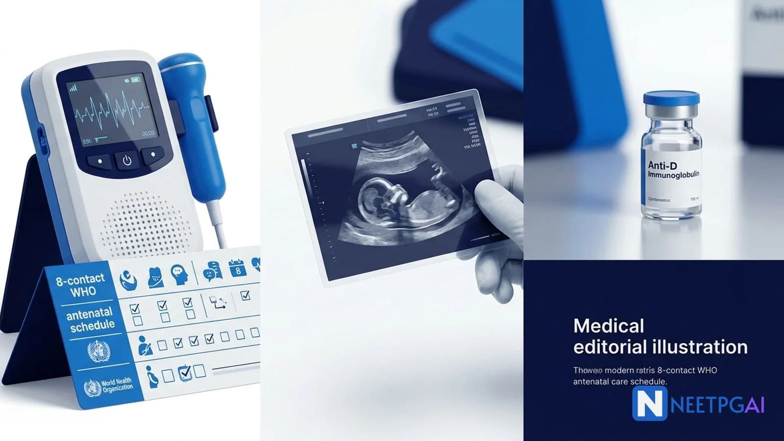
Antenatal Care & Pregnancy Complications for NEET PG — Complete Guide 2026
Master antenatal care and pregnancy complications for NEET PG 2026: WHO 2016 8-contact ANC schedule, booking visit investigations, anomaly scan, DIPSI GDM screening, Rh-negative pregnancy with anti-D, APH comparison, preterm labor tocolytics, PROM vs PPROM, and multiple pregnancy.
NEETPGAI EditorialPublished 31 Jan 202621 min read
Share this article
This content is for educational purposes for NEET PG exam preparation. It is not a substitute for professional medical advice, diagnosis, or treatment. Clinical information has been reviewed by qualified medical professionals.
Ready to put this into practice?
Start practicing NEET PG MCQs with AI-powered explanations.
Antenatal corticosteroids — betamethasone 12 mg IM × 2 doses (24 h apart) at 24–34 weeks; reduce RDS, IVH, NEC
Multiple pregnancy — chorionicity by T1 US (lambda vs T sign); MCMA highest risk, dichorionic lowest; TTTS, IUGR, preterm labor, PPH
Antenatal care is a structured programme of supervision throughout pregnancy that reduces maternal and perinatal mortality — and it is a NEET PG goldmine because it spans obstetrics, community medicine, and public health. The student who memorises the WHO 2016 schedule, DIPSI GDM screening, anti-D timing, and APH differentials covers 4–5 marks across papers. Pair this guide with daily MCQ practice on the obstetrics and gynaecology subject hub, cross-reference the OBG high-yield topics overview, and revise the preeclampsia and eclampsia guide for hypertensive complications integration.
ANC schedule — WHO 2016 vs RMNCH+A
Antenatal care is the systematic medical supervision of a pregnant woman from conception to labour onset, and the WHO 2016 model marks a major shift from the older 4-visit Focused ANC.
WHO 2016 — 8 contacts (the new standard):
Contact
Gestational age
Focus
1
<12 weeks
Booking, dating, risk stratification, baseline labs, folic acid 400 mcg
2
20 weeks
Anomaly scan, anaemia reassessment
3
26 weeks
GDM screen (DIPSI / IADPSG), BP trend
4
30 weeks
Iron/folic acid/calcium compliance, fetal growth
5
34 weeks
Presentation, preeclampsia surveillance, anti-D if Rh-negative
6
36 weeks
Presentation confirmation, birth plan
7
38 weeks
Labour readiness
8
40 weeks
Post-dates plan, membrane sweep if appropriate
Rationale for 8 over 4: the 2016 WHO Cochrane review found the 4-visit FANC model was associated with a small increase in perinatal mortality, driving the evidence-based shift.
India RMNCH+A (minimum 4 visits — operational reality):
Visit 1 — within first trimester (ideally <12 weeks)
Visit 2 — at 14–26 weeks
Visit 3 — at 28–34 weeks
Visit 4 — at 36 weeks to term
Ideal Indian best-practice now recommends 8 contacts aligning with WHO 2016, but field services still often deliver 4. Know both for NEET PG.
Universal supplementation under RMNCH+A:
Folic acid 400 mcg daily from preconception (ideally 3 months before) through first trimester to prevent NTDs; 5 mg if prior NTD child / anticonvulsants
Iron: 60 mg elemental iron + 500 mcg folic acid daily from the 4th month for 180 days, then 180 days postpartum (total 360 days)
Calcium: 1 g daily from the 4th month through pregnancy and 6 months postpartum
Tetanus toxoid / Td: 2 doses 4 weeks apart in first pregnancy; 1 booster in subsequent
Booking visit investigations
The booking (first antenatal) visit is the foundation of antenatal risk stratification — ideally performed between 8 and 12 weeks.
Medical (HTN, diabetes, thyroid, TB, cardiac, epilepsy, haematological)
Surgical (previous caesarean, myomectomy, other abdominal)
Drug history (anticonvulsants, warfarin, ACEi)
Family history (twinning, genetic disorders, hereditary cancers, haemoglobinopathies)
Social (smoking, alcohol, substance use, occupation, partner HIV status)
Examination:
Height, weight, BMI calculation
Vitals (BP in both arms if first antenatal; baseline)
General exam (pallor, oedema, thyroid, cardiovascular)
Obstetric exam (fundal height if gestation >=12 weeks, presentation only in T3)
Mandatory labs at booking:
Investigation
Purpose
CBC
Baseline Hb; thrombocytopenia; MCV for thalassaemia screen
Blood group + Rh
Plan anti-D if Rh-negative
Urine routine + albumin + glucose
UTI, proteinuria baseline
Urine culture
Asymptomatic bacteriuria (treat to prevent pyelonephritis / preterm labour)
Random blood sugar / fasting
Baseline diabetic screen
HIV (opt-out)
Initiate lifelong ART if positive (Option B+)
VDRL / RPR
Syphilis (treat with benzathine penicillin)
HBsAg
Infant HBIG + HBV vaccine within 12 h of birth
HCV
Emerging; in high-risk populations
TSH
Subclinical hypothyroidism (target TSH <2.5 in T1)
Dating ultrasound (11–14 weeks)
EDD confirmation, viability, number, NT measurement for aneuploidy screen
Additional (risk-based):
OGTT 75 g at booking if high-risk (BMI >30, prior GDM, PCOS, family history T2DM, previous macrosomia or stillbirth)
Haemoglobin electrophoresis (thalassaemia / sickle cell) — all in India given high carrier prevalence
Rubella IgG — immunity; do NOT vaccinate in pregnancy
Varicella IgG — if history unclear
TORCH screen only if clinically indicated (not universal)
Anomaly scan — 18–22 weeks
The fetal anomaly scan (Targeted Imaging for Fetal Anomalies, TIFFA) is a detailed level-II ultrasound performed between 18 and 22 weeks that screens for major structural abnormalities.
Indian standard: 18–20 weeks (practical, within PC-PNDT Act compliance; sex determination prohibited and not communicated).
Systematic assessment:
Head and brain — biparietal diameter, head circumference, ventricles (lateral ventricle <=10 mm), cerebellum, cisterna magna, nuchal fold (<6 mm), face (lips/palate/profile)
Spine — longitudinal and transverse views for neural tube defects
Heart — four-chamber view, outflow tracts (LVOT, RVOT), three-vessel view, crossing of great vessels
Placenta — location (rule out previa), cord insertion, amniotic fluid index (AFI 8–18 cm)
Cervix — transvaginal for short cervix (<25 mm) in high-risk preterm labour
Soft markers for aneuploidy (Down syndrome):
Nasal bone absence / hypoplasia (strongest marker)
Increased nuchal fold (>=6 mm at 15–20 weeks)
Echogenic intracardiac focus
Pyelectasis (renal pelvis 4–10 mm)
Short femur / humerus
Echogenic bowel
Single umbilical artery
Choroid plexus cysts (trisomy 18 marker)
Non-invasive prenatal testing (NIPT) screens cell-free fetal DNA in maternal blood from 10 weeks onwards — high sensitivity for T21, T18, T13. Increasingly available but not universal in India.
Delivery — induce at 39–40 weeks for well-controlled diet-treated GDM; 38–39 weeks for insulin-treated
Maternal risks: preeclampsia, polyhydramnios, operative delivery, T2DM later life.
Fetal risks: macrosomia, shoulder dystocia, birth trauma, NICU admission, neonatal hypoglycaemia, hypocalcaemia, jaundice, RDS, stillbirth.
Post-delivery: repeat 75 g OGTT at 6 weeks to identify persistent diabetes; lifelong annual screening.
Rh-negative pregnancy — anti-D and Kleihauer-Betke
Rh-negative pregnancy carries the risk of maternal alloimmunisation to fetal Rh-positive cells, causing haemolytic disease of the fetus and newborn (HDFN) — prevented by anti-D immunoglobulin.
Indian Rh-negative prevalence: ~5–10%, substantially lower than Western populations (~15%).
Pathophysiology:
Fetomaternal haemorrhage (FMH) → fetal Rh-positive RBCs in maternal circulation
Dose of anti-D: 10 mcg covers 0.5 mL of fetal RBCs (1 mL of fetal blood)
If FMH >30 mL, additional 300 mcg per 30 mL of fetal blood
Used when massive FMH suspected (abruption, trauma, stillbirth)
Flow cytometry is an alternative quantitative test.
Antepartum haemorrhage — previa vs abruption vs vasa previa
Antepartum haemorrhage (APH) is bleeding from the genital tract after 20 weeks and before delivery — and the differentiation of previa, abruption, and vasa previa is a classic NEET PG table.
Feature
Placenta previa
Abruptio placentae
Vasa previa
Bleeding
Painless, recurrent, fresh red
Painful, continuous, dark red (or concealed)
Painless, sudden at ROM, fetal blood
Onset
Usually T3, spontaneous
Often T3, may be trauma/HTN-triggered
At rupture of membranes
Cause
Low implantation
Premature separation
Fetal vessels crossing cervix
Uterus
Soft, relaxed
Tender, hypertonic, woody-hard
Soft
Fetal heart
Usually normal
Distress or demise
Rapid fetal exsanguination
DIC risk
Low
High (10–20%)
Low for mother
Ultrasound
Diagnostic — placenta near/covering os
Often falsely negative; retroplacental clot in ~25%
Often diagnostic with colour Doppler
Apt test
Negative
Negative
Positive (fetal Hb alkali-resistant)
Digital PV
Contraindicated until previa excluded
May be done if needed
Avoid
Delivery mode
LSCS if major, especially grade III–IV
Immediate LSCS if fetal distress or maternal compromise; vaginal if IUD and stable
IV bolus 6.75 mg, then 300 mcg/min × 3 hours, then 100 mcg/min up to 48 hours
Cleanest side-effect profile
Expensive — not routine in Indian public sector
Older agents (less preferred):
Ritodrine / Terbutaline (beta-agonists): tachycardia, maternal pulmonary oedema (especially with multiple pregnancy, infection, fluid overload), hyperglycaemia, hypokalaemia. FDA black-box for terbutaline (not for prolonged use)
Magnesium sulfate — less effective as tocolytic, but used for fetal neuroprotection
Indomethacin (COX inhibitor) — effective but premature ductal closure if >=32 weeks; oligohydramnios; used <32 weeks short-term only
Contraindications to tocolysis:
Gestation >=34 weeks
Intrauterine fetal death
Lethal fetal anomaly
Severe preeclampsia / eclampsia
Placental abruption with maternal or fetal compromise
Chorioamnionitis
Non-reassuring fetal status
Advanced cervical dilatation (>=5 cm)
Antenatal corticosteroids (ACS):
Betamethasone 12 mg IM × 2 doses 24 hours apart OR
Dexamethasone 6 mg IM × 4 doses 12 hours apart
Window: 24–34 weeks (some protocols extend to 36+6 weeks for late preterm rescue course)
Benefits: ~40% reduction in RDS, ~50% reduction in IVH, reduced NEC, reduced neonatal mortality
Single rescue course if >7 days since first and still <34 weeks
Magnesium sulfate for fetal neuroprotection:
4 g IV load, then 1 g/h infusion × up to 24 hours
If imminent preterm delivery <32 weeks
Reduces cerebral palsy incidence
GBS prophylaxis:
Penicillin / ampicillin IV at onset of labour if GBS+ / unknown + risk factors
Preterm labour is a risk factor
PROM vs PPROM
PROM (Premature Rupture of Membranes) is rupture of membranes before the onset of labour at term (>=37 weeks); PPROM (Preterm PROM) is membrane rupture before 37 weeks.
Diagnosis:
History of gush of fluid
Pooling of clear fluid in posterior fornix on sterile speculum exam (avoid digital exam to reduce infection)
Nitrazine paper test: amniotic fluid pH >6.5 turns paper blue (false positive with blood, semen, BV, urine)
Fern test: dried fluid shows fern-like crystals under microscope
AmniSure / PAMG-1 — biochemical test
Ultrasound: oligohydramnios (AFI reduced)
Management of PROM at term:
Induction of labour (usually within 24 hours) reduces chorioamnionitis risk
Deliver at 34 weeks OR earlier if chorioamnionitis, abruption, fetal distress, or active labour
Chorioamnionitis signs:
Maternal fever >=38°C
Fetal tachycardia >160
Maternal tachycardia >=100
Uterine tenderness
Foul-smelling vaginal discharge
Maternal leukocytosis (not reliable after steroids)
Multiple pregnancy
Multiple pregnancy is the simultaneous gestation of two or more fetuses — chorionicity rather than zygosity is the single most important antenatal determinant of risk.
Types by zygosity:
Monozygotic (identical) — 1/3 of twins worldwide; fixed rate (~3.5/1000)
Dizygotic (fraternal) — 2/3; rate varies with maternal age, parity, ART, race (highest in Nigeria)
Types by chorionicity (first-trimester US, 11–14 weeks):
Type
Timing of zygote division
Chorionicity
Amnionicity
Incidence in MZ
Risk
DCDA
0–3 days
Dichorionic
Diamniotic
~30%
Lowest (similar to singleton)
MCDA
4–8 days
Monochorionic
Diamniotic
~65%
TTTS, IUGR
MCMA
9–12 days
Monochorionic
Monoamniotic
~1%
Cord entanglement — highest risk
Conjoined
>13 days
Monochorionic
Monoamniotic
<1%
Surgical separation if possible
Dizygotic twins are ALWAYS dichorionic diamniotic.
Ultrasound chorionicity signs (first trimester):
Lambda sign (λ) / twin peak — dichorionic
T sign — monochorionic
Antenatal management:
Dating in first trimester with chorionicity determination
Every 4 weeks for DCDA, every 2 weeks for MCDA, weekly in T3 for MCMA
Screening for preeclampsia, anaemia, GDM, preterm labour
Specific complications:
Twin-to-twin transfusion syndrome (TTTS) — MC twins; shared placental anastomoses; donor (oligohydramnios + small) and recipient (polyhydramnios + large + cardiac failure); treatment = fetoscopic laser ablation at 16–26 weeks
Twin anaemia polycythaemia sequence (TAPS)
Selective IUGR
Single twin demise (vanishing twin; increased risk of neurological injury in surviving MC twin)
Delivery timing (uncomplicated):
DCDA — 38 weeks
MCDA — 36 weeks
MCMA — 32–34 weeks with planned caesarean
Conjoined — caesarean
Mode of delivery (presentation-based):
Both cephalic: vaginal trial
First cephalic, second non-cephalic: vaginal with internal podalic version of second OR LSCS
World Health Organization. WHO Recommendations on Antenatal Care for a Positive Pregnancy Experience (2016 update, reaffirmed 2022).
Government of India, Ministry of Health & Family Welfare. RMNCH+A Guidelines for Antenatal Care and Skilled Attendance at Birth by ANMs/LHVs/SNs (2016 update, 2020 reaffirmation).
Diabetes in Pregnancy Study Group India (DIPSI). Consensus Guidelines on Screening and Diagnosis of GDM (2020 revision).
Williams Obstetrics, 26th Edition (Cunningham, Leveno, Bloom et al., 2022).
DC Dutta's Textbook of Obstetrics, 9th Edition (2018) — Chapters on Antenatal Care, APH, Preterm Labour, Multiple Pregnancy, Rh Isoimmunisation.
Kenyon SL et al. (ORACLE II Collaborative Group). Broad-spectrum antibiotics for preterm, prelabour rupture of fetal membranes: the ORACLE I randomised trial. Lancet 2001; 357:979-988.
Royal College of Obstetricians and Gynaecologists (RCOG). Green-top Guideline No. 27: Placenta Praevia, Placenta Praevia Accreta and Vasa Praevia (2018).
Frequently asked questions
How many ANC and pregnancy complication questions appear in NEET PG?
Antenatal care and pregnancy complications contribute 4-5 direct questions per NEET PG paper across obstetrics, community medicine, and pharmacology. WHO 2016 ANC schedule, anti-D timing in Rh-negative pregnancy, DIPSI GDM screening, APH differentials, and tocolytic choice are the most tested subtopics based on 2019-2025 pattern analysis.
What is the WHO 2016 antenatal care schedule?
WHO 2016 recommends 8 antenatal contacts (a shift from the older 4-visit Focused ANC model) because 4 visits were associated with higher perinatal mortality. The revised schedule: 1 contact in first trimester (before 12 weeks), 2 contacts in second trimester (at 20 and 26 weeks), and 5 contacts in third trimester (at 30, 34, 36, 38, and 40 weeks). India still operates a minimum 4-visit schedule under RMNCH+A, but the 8-contact model is the international standard.
What investigations are done at the booking visit?
Booking visit (ideally at 8-12 weeks) includes complete history and exam, BP, height/weight, CBC, blood group and Rh typing, urine routine and albumin, HIV (opt-out), VDRL, HBsAg, random blood sugar, thyroid screen (TSH), and dating ultrasound. Baseline OGTT is done if high-risk (BMI over 30, prior GDM, prior macrosomia, PCOS, family history). An optional rubella IgG and a baseline ECG are added in select protocols.
When is the anomaly scan done and what does it look for?
The fetal anomaly scan is done between 18 and 22 weeks of gestation (Indian standard is 18-20 weeks under the PC-PNDT Act). It assesses major structural anomalies of the head, spine, heart (four-chamber view, outflow tracts), abdomen, kidneys, and limbs. Markers like nasal bone absence, increased nuchal fold, echogenic intracardiac focus, and ventriculomegaly prompt karyotyping. Earlier scans (11-14 weeks, NT scan) screen for aneuploidy but miss most structural anomalies.
What is DIPSI and how is GDM screened?
DIPSI (Diabetes in Pregnancy Study Group India) recommends universal GDM screening with a 75 g oral glucose challenge regardless of fasting status, with a single venous plasma sample at 2 hours. A value greater than or equal to 140 mg/dL is diagnostic of GDM. DIPSI is practical for low-resource settings because it does not require fasting. Screening is done once at 24-28 weeks (and at booking if high-risk). Alternative: IADPSG 75 g OGTT with fasting, 1-hour, and 2-hour samples (fasting greater than or equal to 92, 1-hour greater than or equal to 180, 2-hour greater than or equal to 153 — any one diagnostic).
When is anti-D given in Rh-negative pregnancy?
Anti-D immunoglobulin 300 micrograms IM is given to Rh-negative unsensitized women at 28 weeks (antenatal prophylaxis) and within 72 hours postpartum if the baby is Rh-positive. Additional doses after any sensitizing event: spontaneous or induced abortion, ectopic pregnancy, antepartum hemorrhage, external cephalic version, amniocentesis, chorionic villus sampling, or abdominal trauma. Kleihauer-Betke test quantifies fetomaternal hemorrhage to decide if more than the standard 300 micrograms is needed.
What is the Kleihauer-Betke test?
The Kleihauer-Betke (acid elution) test detects fetal red blood cells in maternal circulation and quantifies fetomaternal hemorrhage. Maternal blood smear is treated with acid buffer — adult haemoglobin (HbA) elutes leaving ghost cells, while fetal haemoglobin (HbF) is acid-resistant and stains pink. The percentage of fetal cells guides additional anti-D dosing beyond the standard 300 micrograms (each additional 30 mL of fetomaternal bleed needs one more 300 microgram dose).
How do you differentiate placenta previa, abruption, and vasa previa?
Placenta previa is painless, causeless, recurrent, fresh bleeding with a soft relaxed uterus and normal fetal heart sounds — ultrasound shows placental edge less than 2 cm from the internal os. Abruption is painful, dark bleeding with a tender hypertonic uterus, fetal distress or demise, and possible DIC — ultrasound is often falsely negative. Vasa previa is painless bleeding at rupture of membranes with rapid fetal distress or demise (Apt test detects fetal blood).
What is the first-line tocolytic for preterm labor?
Nifedipine is first-line for preterm labor tocolysis in most contemporary guidelines because of oral availability, efficacy, low maternal side effects, and no link to neonatal pulmonary oedema (seen with beta-agonists). Loading dose 20 mg oral, then 10-20 mg every 4-8 hours. Atosiban (oxytocin receptor antagonist) is an equally efficacious alternative with the cleanest side-effect profile but is expensive. Ritodrine and terbutaline (beta-agonists) are older agents with more maternal and fetal side effects.
What is the difference between PROM and PPROM?
PROM (Premature Rupture of Membranes) is rupture of membranes before the onset of labor at term (at or after 37 weeks). PPROM (Preterm PROM) is rupture of membranes before 37 weeks. Diagnosis relies on visual pooling of liquor in the posterior fornix, positive nitrazine test (pH more than 6.5 — blue colour change), positive fern test, and ultrasound showing oligohydramnios. PPROM management includes corticosteroids for lung maturity (24-34 weeks), broad-spectrum antibiotics (erythromycin) for latency, and delivery at 34 weeks or earlier if chorioamnionitis, abruption, or fetal distress occurs.
Explore our pricing plans for unlimited practice across all 19 subjects, AI-powered doubt resolution, and personalized study plans.
This content is for educational purposes for NEET PG exam preparation. It is not a substitute for professional medical advice, diagnosis, or treatment. Clinical information has been reviewed by qualified medical professionals.
Written by: NEETPGAI Editorial Team
Reviewed by: Pending SME Review
Last reviewed: March 2026
This article is reviewed by qualified medical professionals for clinical accuracy and exam relevance. For corrections or updates, contact the editorial team.