Version 1.0 — Published February 2026
Quick Answer
Systemic lupus erythematosus (SLE) is a multisystem autoimmune disease driven by anti-nuclear antibodies and immune-complex deposition, with prevalence of about 30-50 per 100,000 in India and a 9:1 female predominance in reproductive age. In a 24-year-old woman with 3-month symmetric polyarthritis, photosensitive malar rash, oral ulcers, alopecia, fatigue, Hb 9.8 g/dL, thrombocytopenia, 2+ proteinuria, ANA 1:1280 speckled, and anti-dsDNA positive, follow this 5-step workflow:
- Apply ACR/EULAR 2019 classification — ANA positive is the entry criterion; additive weighted points from clinical and immunologic domains; score of 10 or above classifies as SLE
- Screen for lupus nephritis — urinalysis (casts, dysmorphic RBCs), spot urine protein:creatinine ratio, serum creatinine and eGFR; proteinuria above 0.5 g/day or active sediment mandates renal biopsy
- Start hydroxychloroquine 5 mg/kg/day in every SLE patient — reduces flares by 50 percent, improves 10-year survival, lowers thrombosis risk
- Treat proliferative lupus nephritis (Class III or IV on biopsy) with pulse methylprednisolone 500-1000 mg IV × 3 days + oral prednisolone taper + MMF 2-3 g/day (preferred for young women) OR IV cyclophosphamide (Euro-Lupus regimen 500 mg every 2 weeks × 6)
- Plan for pregnancy at least 6 months into stable remission — continue HCQ and azathioprine, stop MMF 6 weeks before conception, add low-dose aspirin from 12 weeks, screen for anti-Ro (fetal heart block risk)
The case
A 24-year-old woman presents to the rheumatology outpatient department with a 3-month history of symmetric, progressive polyarthralgia and morning stiffness lasting 90 minutes. She reports that small joints of both hands (MCPs, PIPs), wrists, and knees are painful and mildly swollen but non-deforming. Over the last 6 weeks she has noticed a red rash across both cheeks and the bridge of her nose that sparingly involves the nasolabial folds and worsens dramatically after 10-15 minutes of sun exposure. For the past month she has had multiple painless oral ulcers on the hard palate, generalized fatigue, a 4-kg unintentional weight loss, and patchy hair thinning from the frontal scalp without visible scarring. She denies fever, chest pain, shortness of breath, abdominal pain, focal neurological deficits, Raynaud phenomenon, or dry eyes/mouth.
She is unmarried, works as a software engineer, has no significant past medical history, no previous pregnancies, and no family history of autoimmune disease. She is on no regular medications, does not smoke, and drinks alcohol only socially. Menstrual cycles are regular; last menstrual period was 10 days ago.
On initial evaluation, her vitals are: pulse 88 bpm regular, BP 128/82 mmHg (mildly raised for her age), respiratory rate 16/min, SpO2 99 percent on room air, temperature 37.3 C axillary. She appears tired and mildly pale but is alert and oriented. Skin exam reveals a fixed, non-pruritic erythematous macular rash over both malar eminences and bridge of nose sparing the nasolabial folds (classic "butterfly" rash). Two shallow ulcers are visible on the hard palate. Scalp shows patchy, non-scarring alopecia over frontal and parietal areas. Joint exam: bilateral symmetric synovitis of 2nd and 3rd MCPs, PIPs, wrists, and knees with no deformity, no erosions clinically.
History and examination
Systemic lupus erythematosus is an autoimmune disease driven by loss of tolerance to nuclear antigens, with formation of immune complexes that deposit in skin, joints, kidneys, serosal surfaces, and the central nervous system. In India, SLE prevalence is estimated at 30-50 per 100,000 population (ICMR multicenter studies 2014-2022), with peak incidence between 15 and 45 years of age and a 9:1 female-to-male ratio. Indian patients are more likely than European cohorts to develop lupus nephritis (40-60 percent vs 25-40 percent) and to present with more severe organ involvement at diagnosis — early referral to rheumatology and proactive renal screening are critical.
General examination:
- Alert, oriented, mildly fatigued
- Pallor (conjunctival, palmar creases) consistent with anemia
- No icterus, no lymphadenopathy, no pedal edema at this visit
- Weight 52 kg, height 158 cm, BMI 20.8 kg/m2
Skin and mucosal examination:
- Classic malar (butterfly) rash — fixed erythema over both malar eminences and nose bridge, sparing the nasolabial folds (helps distinguish from rosacea or seborrheic dermatitis)
- Photosensitivity reported (worsens with 10-15 minutes of sun exposure)
- Multiple shallow, painless oral ulcers on the hard palate (painless is the clue — unlike aphthous ulcers which are painful)
- Patchy non-scarring alopecia on the scalp (active disease pattern; discoid lupus produces scarring alopecia — different entity)
- No discoid lesions, no livedo reticularis, no Raynaud discoloration noted
Musculoskeletal examination:
- Symmetric synovitis of 2nd and 3rd MCPs and PIPs bilaterally, both wrists, both knees
- Grip weakness mildly reduced from joint pain
- No deformity, no ulnar deviation, no swan-neck or boutonniere changes (SLE arthritis is classically non-erosive and non-deforming — Jaccoud arthropathy may develop with chronic disease)
- No muscle weakness or tenderness
Cardiopulmonary examination:
- Heart sounds normal, no murmur, no pericardial rub
- Chest clear to auscultation, no pleural rub
- No evidence of active serositis today
Abdominal and neurological examination:
- Abdomen soft, non-tender, no organomegaly
- Neurological examination grossly normal (GCS 15, no focal deficit, no meningism)
- No psychiatric features on screening
Differential diagnosis
Systemic lupus erythematosus is the leading diagnosis given the multisystem involvement and photosensitive rash, but several connective tissue and non-autoimmune conditions must be considered and systematically excluded.
| Diagnosis | Points in favor | Points against |
|---|
| Systemic lupus erythematosus (SLE) | Multisystem: skin, joints, mucosa, hematologic, renal; photosensitive malar rash; oral ulcers; non-erosive arthritis | None — all features fit |
| Rheumatoid arthritis | Symmetric polyarthritis of small hand joints with morning stiffness | No malar rash, no oral ulcers, no photosensitivity, no renal or mucocutaneous involvement; RF and anti-CCP would be needed to confirm if atypical |
| Sjogren syndrome | Female, connective tissue features, ANA positive possible | No sicca symptoms (dry eyes, dry mouth), no parotid enlargement; Schirmer test needed |
| Mixed connective tissue disease (MCTD) | Female, connective tissue overlap features | Raynaud phenomenon typically prominent; anti-U1-RNP in very high titer required |
| Dermatomyositis | Female, rash, fatigue | No heliotrope rash, no Gottron papules, no proximal muscle weakness, no raised CK |
| Rosacea / seborrheic dermatitis | Facial erythema | No systemic features, no joint, mucosal, or renal involvement; rosacea involves nasolabial folds, SLE spares them |
| Viral polyarthritis (parvovirus B19, chikungunya, dengue-associated) | Acute symmetric polyarthritis | No preceding fever, no rash consistent with viral exanthem, 3-month duration too long for typical viral arthritis |
| Drug-induced lupus | SLE-like features possible | Not on any offending drug (hydralazine, procainamide, minocycline, isoniazid, anti-TNF agents); anti-histone antibodies would be expected |
The combination of symmetric non-erosive polyarthritis, photosensitive malar rash sparing nasolabial folds, painless oral ulcers on hard palate, non-scarring alopecia, anemia, thrombocytopenia, proteinuria, and a positive ANA with high-titer anti-dsDNA is pathognomonic for systemic lupus erythematosus with probable lupus nephritis — renal biopsy is the next step.
Investigations
Initial investigations aim to confirm SLE by ACR/EULAR 2019 criteria, identify active organ involvement (especially renal), and establish baseline for monitoring.
- Complete blood count: Hb 9.8 g/dL (normocytic normochromic anemia of chronic disease; Coombs test to rule out AIHA), WBC 3,400 (mild leukopenia), lymphocytes 1,100 (lymphopenia < 1500 is an SLE criterion), platelets 88,000 (thrombocytopenia < 100,000 is an SLE criterion)
- Direct Coombs test: negative (rules out autoimmune hemolytic anemia; anemia here is chronic disease pattern)
- ESR / CRP: ESR 68 mm/hr (raised); CRP 6 mg/L (mildly raised — disproportionate ESR to CRP is classic for SLE; high CRP suggests concurrent infection or serositis)
- Renal panel: creatinine 0.9 mg/dL, urea 28 mg/dL, eGFR 92 mL/min (preserved GFR despite proteinuria — early nephritis)
- Urinalysis: 2+ proteinuria, 10-15 dysmorphic RBCs per HPF, occasional red cell casts — active urinary sediment, strongly suggestive of glomerulonephritis
- Spot urine protein:creatinine ratio: 1.2 g/g (= 1.2 g/day proteinuria — above 0.5 g/day threshold mandates renal biopsy)
- Liver function tests: within normal limits
- Complement C3 and C4: C3 62 mg/dL (low; normal 90-180), C4 8 mg/dL (low; normal 10-40) — low complement indicates active disease with consumption, classic for lupus nephritis
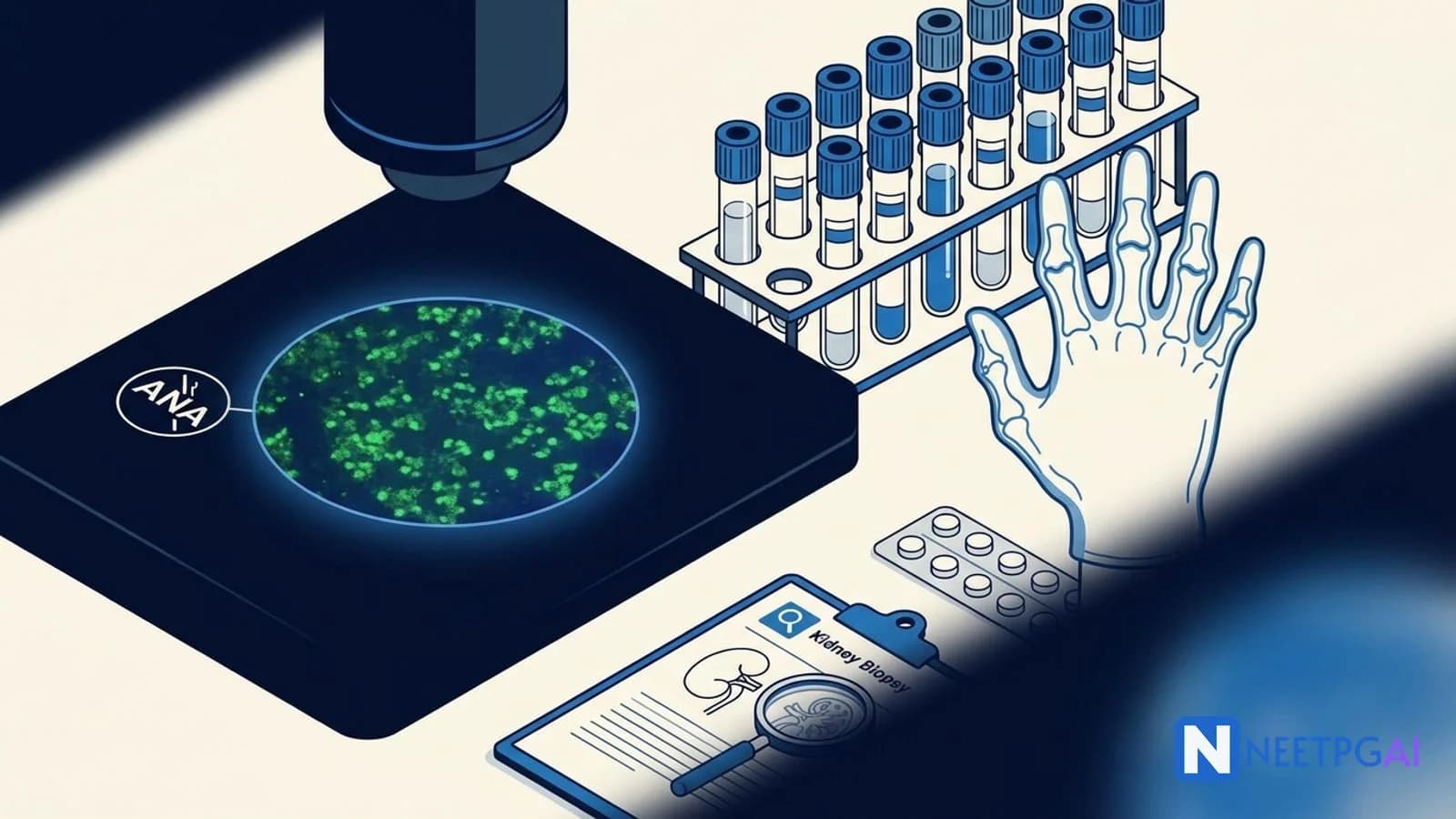
- ANA by indirect immunofluorescence on HEp-2 cells: positive at 1:1280 titer with speckled pattern (entry criterion for ACR/EULAR 2019 satisfied)
- Anti-dsDNA: positive at high titer (specific for SLE; correlates with disease activity and nephritis)
- Anti-Smith (anti-Sm): positive (highly specific for SLE — nearly 99 percent specificity, though only 25-30 percent sensitivity)
- Anti-Ro/SSA and anti-La/SSB: anti-Ro positive, anti-La negative (relevant for future pregnancy planning — congenital complete heart block risk)
- Anti-RNP, anti-Scl-70, anti-Jo-1, anti-centromere: negative (rules out MCTD, systemic sclerosis, myositis)
- Antiphospholipid antibody panel: lupus anticoagulant negative, anti-cardiolipin IgG/IgM negative, anti-beta2-glycoprotein-I negative (no antiphospholipid syndrome today — repeat in 12 weeks if initial suspicion)
- Renal biopsy (ultrasound-guided percutaneous, after platelet correction): diffuse proliferative glomerulonephritis with endocapillary hypercellularity involving more than 50 percent of glomeruli, wire-loop lesions, fibrinoid necrosis, and full-house immunofluorescence (IgG, IgA, IgM, C3, C4, C1q deposits) — Class IV (A) lupus nephritis per ISN/RPS 2003 classification
ACR/EULAR 2019 classification scoring for this patient:
| Domain | Item | Weight |
|---|
| Entry | ANA 1:1280 speckled | Satisfies entry |
| Mucocutaneous | Oral ulcers | 2 |
| Mucocutaneous | Non-scarring alopecia | 2 |
| Mucocutaneous | Acute cutaneous lupus (malar rash) | 6 |
| Musculoskeletal | Joint involvement (synovitis in 2 or more joints) | 6 |
| Hematologic | Thrombocytopenia < 100,000 | 4 |
| Hematologic | Leukopenia < 4,000 | 3 |
| Renal | Class III or IV lupus nephritis on biopsy | 10 |
| Immunologic (complement) | Low C3 AND low C4 | 4 |
| Immunologic (antibody) | Anti-dsDNA positive OR anti-Sm positive | 6 |
| Total (highest per domain) | Mucocutaneous 6 + MSK 6 + Heme 4 + Renal 10 + Complement 4 + Antibody 6 | 36 points |
Total score is 36 — well above the 10-point threshold. The patient meets ACR/EULAR 2019 classification for SLE with biopsy-proven Class IV lupus nephritis.
Diagnosis
Systemic lupus erythematosus (ACR/EULAR 2019 score 36) with biopsy-proven Class IV (A) diffuse proliferative lupus nephritis, autoimmune cytopenias (mild thrombocytopenia and leukopenia), anemia of chronic disease, acute cutaneous lupus, non-scarring alopecia, oral ulcers, and non-erosive inflammatory polyarthritis in a previously well 24-year-old woman, with:
- High-titer anti-dsDNA and anti-Sm antibodies (SLE-specific)
- Anti-Ro/SSA positive (relevant for future pregnancy planning)
- Low C3 and C4 (active disease)
- Preserved eGFR 92 mL/min with 1.2 g/day proteinuria (active but not yet severe renal impairment — favorable window for induction therapy)
- No antiphospholipid antibodies, no CNS involvement, no serositis, no hemolytic anemia at presentation
Management
Management of newly diagnosed SLE with proliferative lupus nephritis follows four simultaneous streams, aligned with 2024 EULAR and KDIGO guidelines.
Stream 1: Hydroxychloroquine for every SLE patient — start immediately
Hydroxychloroquine (HCQ) 5 mg/kg actual body weight per day (maximum 400 mg/day). For this 52-kg patient: 260 mg/day (rounded to 200 mg once daily, alternating with 300 mg on alternate days — or 200 mg daily given practical dispensing; some centres use 200 mg twice daily for more severe disease then de-escalate).
Five evidence-based benefits:
- Reduces SLE flare rates by 50 percent (LUMINA cohort)
- Improves 10-year survival by 25 percent (Toronto Lupus Cohort)
- Lowers thrombosis risk (relevant given anti-Ro positive)
- Improves lipid profile
- Disease-modifying in lupus nephritis — reduces proteinuria and preserves renal function
Mandatory baseline ophthalmology assessment before starting, then annual screening after 5 years of therapy (earlier with retinal disease, renal impairment, or tamoxifen co-therapy). HCQ retinopathy is the main long-term toxicity but rare at weight-appropriate dosing.
Stream 2: Induction therapy for Class IV lupus nephritis — start within days
2024 EULAR/ACR guidelines recommend the following first-line induction regimen for proliferative lupus nephritis:
Step 1 — Pulse methylprednisolone:
- 500-1000 mg IV daily for 3 consecutive days (lower-end 500 mg pulses are increasingly preferred to reduce steroid toxicity)
Step 2 — Oral prednisolone taper (after pulse):
- Start at 0.5-0.8 mg/kg/day (lower than the traditional 1 mg/kg/day — recent evidence supports steroid-sparing)
- Taper over 3-6 months to maintenance 5-7.5 mg/day
- Give calcium 1000 mg + vitamin D 800 IU daily and consider bisphosphonate if long-term steroid exposure anticipated
Step 3 — Choose one immunosuppressant (MMF preferred here):
| Option | Regimen | Best for |
|---|
| Mycophenolate mofetil (MMF) | 2-3 g/day oral in divided doses × 6 months | Young women of childbearing age (less ovarian toxicity than cyclophosphamide), African/Hispanic patients, moderate-severity Class III/IV |
| Cyclophosphamide (NIH regimen) | 0.5-1 g/m2 IV monthly × 6 months | Severe rapidly progressive nephritis, crescentic disease, non-response to MMF |
| Cyclophosphamide (Euro-Lupus regimen) | 500 mg IV every 2 weeks × 6 doses (total 3 g over 12 weeks) | Preferred in European/Asian patients with milder proliferative nephritis — similar efficacy to NIH regimen, lower cumulative toxicity |
| Belimumab add-on | 10 mg/kg IV at 0, 2, 4 weeks, then monthly | Add to MMF or cyclophosphamide for patients with incomplete response at 3-6 months (BLISS-LN trial showed benefit) |
| Voclosporin add-on | 23.7 mg twice daily (calcineurin inhibitor) | Add to MMF for rapid proteinuria reduction (AURORA trial showed 40 percent complete renal response at 1 year) |
For this patient (24-year-old woman, 52 kg, Class IV(A) lupus nephritis, planning future pregnancy):
- HCQ 200 mg/day (or 260 mg-equivalent weight-based)
- IV methylprednisolone 500 mg daily × 3 days, then oral prednisolone 30 mg/day tapering over 3-6 months
- MMF 2 g/day titrating to 2.5-3 g/day as tolerated × 6 months — MMF is preferred over cyclophosphamide in young women of childbearing age because it spares ovarian function
Stream 3: Adjunctive and supportive therapy
- BP control below 130/80 mmHg — use an ACE inhibitor (ramipril 2.5-10 mg/day) or ARB (telmisartan 40-80 mg/day) for antiproteinuric and renoprotective effect
- Statin for hyperlipidemia from proteinuria and steroids (atorvastatin 10-20 mg/day)
- Pneumococcal, influenza, and SARS-CoV-2 vaccinations (inactivated — administer before heavy immunosuppression if possible; live vaccines contraindicated)
- Bone protection — calcium 1000 mg + vitamin D 800 IU daily; consider bisphosphonate if steroid exposure >= 3 months at 7.5 mg/day or more
- Sunscreen SPF 50+ broad-spectrum and sun-protective clothing — critical for photosensitive skin disease
- Contraception during cytotoxic therapy — MMF is teratogenic; dual barrier + hormonal IUD or progesterone-only methods preferred (estrogen-containing contraceptives acceptable if no antiphospholipid antibodies)
- Smoking cessation — smoking reduces HCQ efficacy and worsens cutaneous disease
Stream 4: Maintenance therapy and long-term monitoring
After 6 months of successful induction (proteinuria below 0.5 g/day, stable eGFR, inactive sediment), switch to maintenance:
| Agent | Dose | Duration |
|---|
| Hydroxychloroquine | Continue 5 mg/kg/day | Lifelong |
| MMF maintenance | 1-2 g/day | At least 3-5 years after remission; longer in high-risk patients |
| Azathioprine | 2 mg/kg/day (alternative to MMF; pregnancy-compatible) | At least 3-5 years |
| Low-dose prednisolone | 2.5-5 mg/day; withdraw if possible after 2 years of stable remission | Minimum necessary |
Monitoring schedule:
| Test | Frequency |
|---|
| CBC, creatinine, LFT, urinalysis, urine protein:creatinine | Every 4-8 weeks during induction; every 3 months in maintenance |
| Complement C3/C4, anti-dsDNA titer | Every 3 months (rising dsDNA with falling complement predicts nephritis flare 2-4 months ahead) |
| SLEDAI-2K score | Every clinic visit |
| Lipid profile, fasting glucose, bone density | Annually |
| Ophthalmology for HCQ retinopathy | Baseline, then annually after 5 years of therapy |
| Cervical cancer and HPV screening | Annually (SLE patients on immunosuppressants have elevated HPV persistence) |
Pregnancy planning in SLE
For this patient, pregnancy can be considered after at least 6 months of stable remission on maintenance therapy. Key rules:
| Action | Timing | Rationale |
|---|
| Discuss contraception | At every visit during cytotoxic therapy | MMF teratogenic; ensure effective contraception |
| Switch from MMF to azathioprine | 6 weeks before conception attempts | MMF teratogenic (cleft lip/palate, microtia, facial dysmorphism) |
| Continue hydroxychloroquine | Throughout pregnancy | Stopping HCQ triples flare rate; HCQ also reduces congenital heart block risk in anti-Ro positive mothers |
| Add low-dose aspirin | From 12 weeks gestation | Reduces preeclampsia risk (universal for SLE pregnancies) |
| Fetal echocardiography | Weekly to fortnightly from 16-26 weeks | Anti-Ro positive — monitor for congenital complete heart block |
| Deliver at tertiary center | Planned | Rheumatology + maternal-fetal medicine co-management |
| Consider LMWH + aspirin | Throughout pregnancy | Only if antiphospholipid syndrome co-exists (not in this patient currently) |
Review the autoimmune connective tissue diseases guide for comparison with Sjogren, systemic sclerosis, and dermatomyositis. Strengthen glomerular disease pattern recognition with the glomerulonephritis complete guide.
How NEET PG tests SLE
SLE contributes 2-3 questions per NEET PG paper across Medicine and Rheumatology, tested through six dominant patterns:
Pattern 1 — The classification criteria question: A vignette gives a patient with multisystem features. You must identify ACR/EULAR 2019 criteria met and decide if classification threshold (10 points) is reached. Trap: forgetting that ANA is the entry criterion — without ANA, no classification regardless of other points.
Pattern 2 — The specific antibody question: Match antibody to disease. Anti-dsDNA and anti-Sm are SLE-specific; anti-Ro/La = Sjogren (also in SLE — fetal heart block risk); anti-Scl-70 = diffuse systemic sclerosis; anti-centromere = limited systemic sclerosis/CREST; anti-Jo-1 = antisynthetase myositis; anti-U1-RNP high titer = MCTD. Trap: remembering ANA as specific — ANA is sensitive but non-specific (positive in 20 percent of healthy elderly and many CTDs).
Pattern 3 — The lupus nephritis biopsy class question: Match ISN/RPS class to biopsy description. Class IV (diffuse, >= 50 percent glomeruli, full-house IF) is commonest and needs aggressive induction. Class V (membranous, subepithelial deposits) presents with nephrotic syndrome. Trap: confusing Class III (focal < 50 percent) and Class IV — both are proliferative and both need induction.
Pattern 4 — The induction therapy question: First-line for Class IV lupus nephritis = HCQ + pulse steroids + MMF (young women) or cyclophosphamide (severe/crescentic). Trap: offering azathioprine as induction — azathioprine is maintenance, not induction.
Pattern 5 — The drug toxicity question: HCQ → retinopathy (annual ophthalmology after 5 years); cyclophosphamide → infertility, hemorrhagic cystitis (give mesna), myelosuppression, bladder cancer long-term; MMF → teratogenicity, GI upset, cytopenias; belimumab → infections. Trap: attributing retinopathy to chloroquine only — hydroxychloroquine also causes it, just less commonly.
Pattern 6 — The pregnancy-in-lupus question: Continue HCQ, switch MMF to azathioprine 6 weeks preconception, add low-dose aspirin from 12 weeks, screen fetal echo if anti-Ro positive. Trap: stopping HCQ during pregnancy (common wrong answer — it must be continued).
High-yield one-liners for last-day revision:
- ACR/EULAR 2019: ANA entry + score 10+ across weighted domains
- Class IV lupus nephritis = diffuse proliferative (>= 50 percent glomeruli); needs induction with MMF or cyclophosphamide
- HCQ for every SLE patient — 5 mg/kg/day, annual eye check after 5 years
- Anti-dsDNA and anti-Smith = SLE-specific; low C3/C4 = active disease
- Pulse methylprednisolone 500-1000 mg IV × 3 days before oral steroids in severe disease
- Euro-Lupus cyclophosphamide = 500 mg every 2 weeks × 6 doses (less toxic than NIH regimen)
- Stop MMF 6 weeks before conception; continue HCQ in pregnancy
- Anti-Ro positive = fetal complete heart block risk — fetal echo 16-26 weeks
- Add low-dose aspirin from 12 weeks for all lupus pregnancies
- SLEDAI-2K for disease activity; SLICC/ACR Damage Index for permanent damage
Frequently asked questions
What are the ACR/EULAR 2019 classification criteria for SLE?
ACR/EULAR 2019 classification requires a positive ANA at a titer of at least 1:80 on HEp-2 cells as an entry criterion — without ANA you cannot classify as SLE. Then additive weighted points from 7 clinical domains (constitutional, hematologic, neuropsychiatric, mucocutaneous, serosal, musculoskeletal, renal) and 3 immunologic domains (antiphospholipid antibodies, complement, SLE-specific antibodies). Each domain contributes the highest-weighted item only. A total score of 10 or above classifies as SLE. The highest-weighted single item is Class III or IV lupus nephritis on biopsy (10 points alone). Criteria are for classification, not diagnosis — clinical judgment still matters in borderline cases. Published jointly by ACR and EULAR in 2019, these criteria have 96 percent sensitivity and 93 percent specificity.
How is lupus nephritis classified and why does it matter?
The ISN/RPS 2003 classification (updated 2018) divides lupus nephritis into six classes based on renal biopsy findings. Class I — minimal mesangial (normal light microscopy, mesangial immune deposits on IF/EM). Class II — mesangial proliferative. Class III — focal (below 50 percent of glomeruli involved). Class IV — diffuse (50 percent or more involved). Class V — membranous (subepithelial deposits, nephrotic syndrome). Class VI — advanced sclerosing (90 percent or more globally sclerosed). Classes III and IV are the proliferative forms — they require aggressive induction immunosuppression with MMF or cyclophosphamide plus high-dose steroids. Class V is treated with MMF plus moderate steroids if nephrotic. Classes I and II rarely need immunosuppression beyond SLE baseline therapy. Biopsy is mandatory for any SLE patient with proteinuria above 0.5 g/day or active urinary sediment.
What is the first-line induction therapy for proliferative lupus nephritis?
First-line induction for Class III or IV lupus nephritis in 2024 EULAR/ACR guidelines is hydroxychloroquine (5 mg/kg/day, always) PLUS pulse methylprednisolone (500-1000 mg IV daily for 3 days) followed by oral prednisolone tapered from 0.5-1 mg/kg/day PLUS either mycophenolate mofetil (MMF 2-3 g/day) or cyclophosphamide (NIH regimen — 0.5-1 g/m2 IV monthly for 6 months; or Euro-Lupus regimen — 500 mg IV every 2 weeks for 6 doses). MMF is preferred for young women of childbearing age (avoids cyclophosphamide ovarian toxicity) and for African and Hispanic patients (better response). Cyclophosphamide is preferred for severe rapidly progressive nephritis and crescentic disease. Belimumab or voclosporin may be added for partial responders. Maintenance after 6 months uses lower-dose MMF (1-2 g/day) or azathioprine (2 mg/kg/day) for at least 3-5 years.
Why is hydroxychloroquine given to every SLE patient?
Hydroxychloroquine (HCQ) is the cornerstone of SLE management and is recommended for every patient regardless of disease activity or organ involvement. Five evidence-based benefits: reduces SLE flare rates by 50 percent (LUMINA cohort), improves 10-year survival (observational studies show 25 percent mortality reduction), lowers thrombosis risk (important in lupus with antiphospholipid antibodies), improves lipid profile, and provides disease-modifying effects in lupus nephritis by reducing proteinuria and preserving GFR. Dose is 5 mg/kg actual body weight per day (maximum 400 mg/day). Annual ophthalmology screening is mandatory after 5 years of therapy (or baseline if retinal risk factors) — HCQ retinopathy is the main long-term toxicity but rare at correct dosing. Stopping HCQ doubles the risk of flares, so compliance must be emphasized.
How is SLE disease activity monitored over time?
SLEDAI-2K (SLE Disease Activity Index 2000) is the standard clinical tool — a weighted score across 24 descriptors covering CNS, vascular, renal, musculoskeletal, serosal, cutaneous, immunologic, and constitutional items. Scores range from 0 to 105. A SLEDAI score of 6 or above indicates clinically meaningful disease activity requiring therapy intensification. Complementary tools include the BILAG 2004 (British Isles Lupus Assessment Group) index for organ-specific scoring and the SLICC/ACR Damage Index (SDI) for permanent damage accumulation. Laboratory monitoring includes complement (C3, C4 — low levels indicate active disease, especially nephritis), anti-dsDNA titer (rising titer correlates with nephritis flare), CBC, creatinine, urinalysis, and spot urine protein:creatinine ratio every 3 months. Annual monitoring: lipid profile, fasting glucose, bone density (steroid exposure), ophthalmology (HCQ).
What medications are safe in lupus pregnancy and what counseling is essential?
Pregnancy planning in SLE requires at least 6 months of stable remission before conception to minimize flare and fetal loss. Safe medications in pregnancy: hydroxychloroquine (continue — discontinuation triples flare rate), azathioprine (up to 2 mg/kg/day), low-dose prednisolone, tacrolimus, cyclosporine, and heparin. Add low-dose aspirin (75-150 mg/day from 12 weeks) for all lupus pregnancies — reduces preeclampsia risk. Contraindicated in pregnancy: mycophenolate mofetil (teratogenic — stop 6 weeks before conception), cyclophosphamide, methotrexate, leflunomide, ACE inhibitors, ARBs, warfarin (first trimester). Anti-Ro/SSA or anti-La/SSB positive patients need fetal echocardiography at 16-26 weeks (congenital complete heart block risk). Antiphospholipid-syndrome co-existence requires LMWH plus aspirin prophylaxis throughout pregnancy. Deliver at a tertiary center with rheumatology and maternal-fetal-medicine support.
How is SLE distinguished from other connective tissue diseases?
Five connective tissue diseases share overlapping features but have distinguishing markers. SLE — multisystem with anti-dsDNA (specific), anti-Smith (highly specific), low complement, lupus nephritis. Sjogren syndrome — sicca symptoms (dry eyes, dry mouth) with anti-Ro/SSA and anti-La/SSB; Schirmer test positive; lip biopsy shows lymphocytic sialadenitis. Systemic sclerosis — skin thickening, Raynaud, esophageal dysmotility, renal crisis; anti-Scl-70 (diffuse), anti-centromere (limited/CREST), anti-RNA polymerase III (renal crisis). Dermatomyositis — heliotrope rash, Gottron papules, shawl sign, proximal myopathy, raised CK; anti-Mi-2, anti-Jo-1 (antisynthetase syndrome). Mixed connective tissue disease (MCTD) — overlap features plus anti-U1-RNP in high titer; Raynaud prominent. ANA is positive in all; the specific antibody panel (dsDNA, Sm, Ro, La, Scl-70, centromere, Jo-1, U1-RNP) drives the diagnosis.
How is SLE tested in NEET PG?
NBE tests SLE through six patterns: ACR/EULAR 2019 classification criteria (entry ANA + additive weighted domains, score 10+ classifies), specific autoantibody matching (anti-dsDNA and anti-Smith are SLE-specific; low complement supports active disease), lupus nephritis biopsy class recognition (Class IV diffuse proliferative is commonest and most aggressive), first-line induction therapy for proliferative nephritis (HCQ + pulse steroids + MMF or cyclophosphamide), drug side effect questions (HCQ retinopathy, cyclophosphamide infertility and hemorrhagic cystitis, MMF teratogenicity), and pregnancy management (stop MMF before conception, continue HCQ, add aspirin, monitor for congenital heart block if anti-Ro positive). Expect 2-3 SLE questions per NEET PG paper in Medicine and Rheumatology sections, often as multistep clinical vignettes.
This content is for educational purposes for NEET PG exam preparation. It is not a substitute for professional medical advice, diagnosis, or treatment. Clinical information has been reviewed by qualified medical professionals.
Sources and references
- Aringer M, Costenbader K, Daikh D, et al. "2019 European League Against Rheumatism/American College of Rheumatology Classification Criteria for Systemic Lupus Erythematosus," Annals of the Rheumatic Diseases, 2019 — the canonical paper defining the ACR/EULAR 2019 criteria with entry ANA requirement and weighted additive scoring.
- Fanouriakis A, Kostopoulou M, Andersen J, et al. "EULAR recommendations for the management of systemic lupus erythematosus: 2023 update," Annals of the Rheumatic Diseases, 2024 — current guidance on HCQ for all, induction regimens for lupus nephritis, and pregnancy care.
- Harrison's Principles of Internal Medicine, 21st Edition (McGraw-Hill, 2022) — Chapter on SLE covers pathogenesis, ISN/RPS lupus nephritis classes, and therapeutic algorithm for Indian clinical practice.
Build strong rheumatology pattern recognition with the medicine subject page, the companion autoimmune connective tissue diseases guide, and the glomerulonephritis complete guide for renal pattern correlation. Ready for unlimited AI-powered MCQs with detailed explanations? Explore NEETPGAI Pro.
For personalized study guidance on rheumatology pattern recognition, try the AI Tutor — it adapts to your weak areas and explains concepts the way a senior resident would.
Written by: NEETPGAI Editorial Team
Reviewed by: Pending SME Review
Last reviewed: February 2026
This article is reviewed by qualified medical professionals for clinical accuracy and exam relevance. For corrections or updates, contact the editorial team.