10 Common Mistakes in Microbiology NEET PG — And How to Avoid Them | NEETPGAI
microbiologymistake guideneet pg 2026
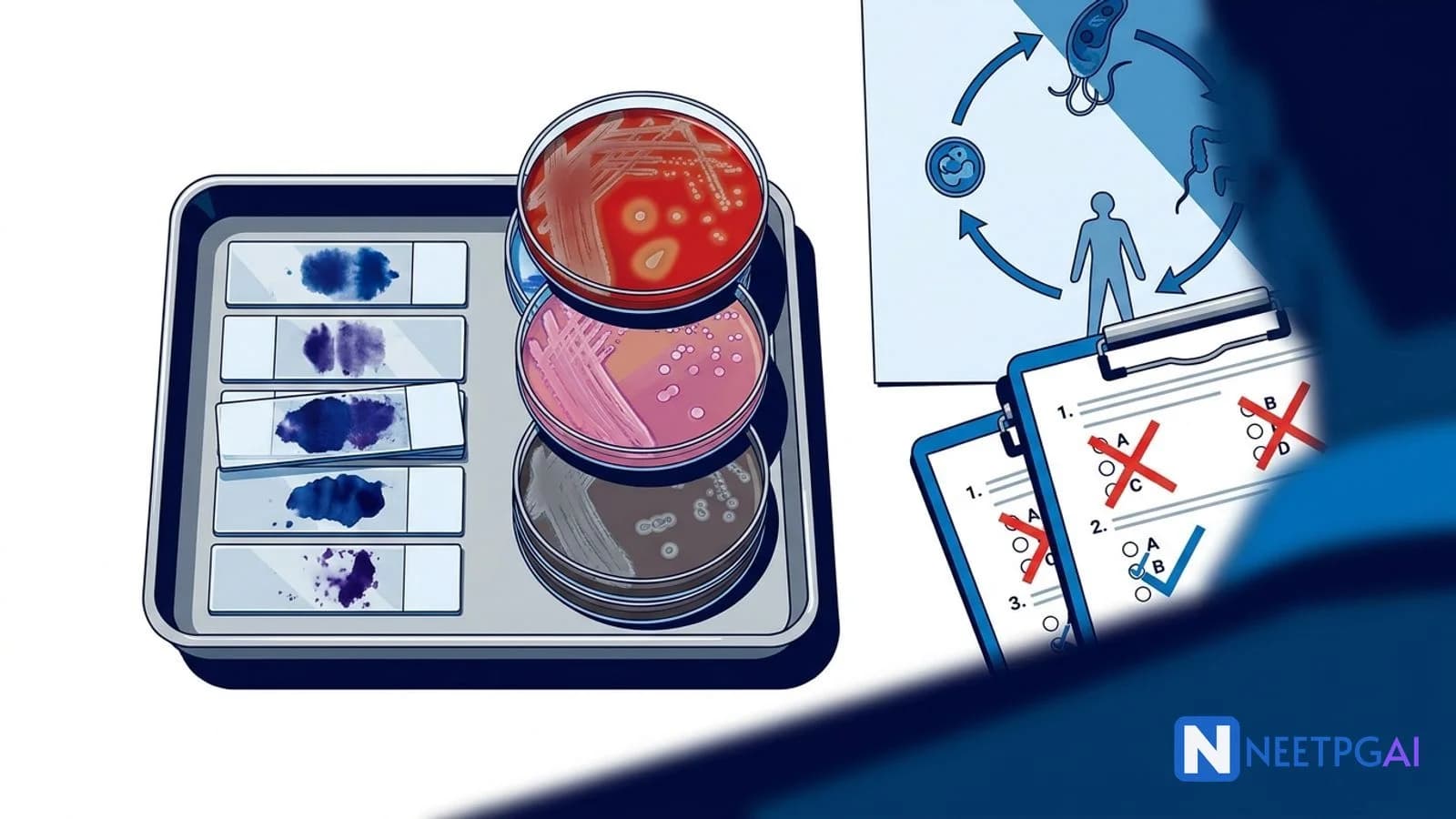
10 Common Mistakes in Microbiology NEET PG — And How to Avoid Them
Avoid the 10 costliest microbiology mistakes in NEET PG 2026: confused gram stain morphology, mixed-up Staph and Strep species, wrong culture media, hepatitis serology traps, parasite lifecycle confusion, antibiotic class mechanisms, dimorphic fungi, virology test selection, bacterial toxin types, and vaccine classification.
NEETPGAI EditorialPublished 15 Jan 202627 min read
Share this article
This content is for educational purposes for NEET PG exam preparation. It is not a substitute for professional medical advice, diagnosis, or treatment. Clinical information has been reviewed by qualified medical professionals.
Ready to put this into practice?
Start practicing NEET PG MCQs with AI-powered explanations.
The single costliest microbiology mistake in NEET PG is confusing similar bacteria within the same genus — mixing up Staph aureus vs epidermidis or Strep pyogenes vs pneumoniae cascades across 2-3 questions per paper. To protect your 15-20 microbiology marks:
Build differentiation tables for each bacterial genus — Staph (coagulase, mannitol), Strep (bacitracin, optochin, bile solubility, quellung reaction), Neisseria (maltose fermentation), Clostridium (toxin-specific diseases)
Group culture media by the organism they select — Lowenstein-Jensen (TB), Loeffler (diphtheria), Thayer-Martin (Neisseria), MacConkey (gram-negative rods, lactose fermenters), Sabouraud (fungi)
Microbiology contributes 15-20 questions to NEET PG, making it the third-highest-weighted pre-clinical subject after pharmacology and pathology (2021-2024 pattern analysis). Unlike pharmacology, which tests mechanism of action, microbiology questions test pattern recognition across morphology, culture characteristics, serology, and clinical correlation. A single conceptual error — for example, confusing Staph aureus with Staph epidermidis — can cost you 2-3 marks in the same paper because the wrong-answer logic propagates.
The ten mistakes below are the patterns that consistently appear in wrong-answer analyses from AIIMS, PGI, and private coaching institutes. Each mistake includes what students typically do, why it fails, the correct approach, and an example MCQ demonstrating the trap.
What students do: Remember only the color (purple = gram positive, pink = gram negative) and forget the shape and arrangement patterns that identify specific organisms.
Why it is wrong: Gram stain gives three pieces of information — color (positive/negative), shape (cocci/bacilli/coccobacilli), and arrangement (clusters, chains, pairs, tetrads). Missing any dimension makes identification impossible. Streptococci are gram-positive cocci in chains. Staphylococci are gram-positive cocci in clusters. Neisseria are gram-negative diplococci. Haemophilus is a gram-negative coccobacillus. A gram stain answer of "gram-positive cocci" without arrangement is 50 percent incomplete.
Correct approach: Memorize the 4-feature gram classification table for the top 20 organisms tested.
Morphology
Arrangement
Classic organisms
Gram-positive cocci
Clusters
Staphylococcus aureus, S. epidermidis, S. saprophyticus
Gram-positive cocci
Chains or pairs
Streptococcus pyogenes, S. pneumoniae, S. agalactiae, Enterococcus
Neisseria meningitidis, N. gonorrhoeae, Moraxella catarrhalis
Gram-negative coccobacilli
Short rods
Haemophilus, Bordetella, Brucella, Pasteurella
Gram-negative bacilli
Enteric rods
Enterobacteriaceae (E. coli, Klebsiella, Salmonella, Shigella, Proteus)
Gram-negative curved/comma
Curved rods
Vibrio cholerae, Campylobacter, Helicobacter
Gram-negative spirochetes
Helical
Treponema pallidum, Leptospira, Borrelia
Example MCQ:A 24-year-old soldier presents with high fever, petechial rash, and meningitis. CSF gram stain shows gram-negative diplococci inside polymorphs. The most likely organism is:
(a) Streptococcus pneumoniae
(b) Neisseria meningitidis
(c) Haemophilus influenzae type b
(d) Listeria monocytogenes
Answer: (b). Gram-negative diplococci inside polymorphs plus meningitis in a young adult with petechial rash is classic meningococcemia. Pneumococcus is gram-positive diplococci; Hib is gram-negative coccobacillus; Listeria is gram-positive bacillus.
Mistake 2: Mixing up similar Staph and Strep species
What students do: Memorize that Staph causes infections and Strep causes infections, without distinguishing the species within each genus.
Why it is wrong: Each species causes different diseases, has different diagnostic features, and responds to different antibiotics. Staph aureus causes TSS, food poisoning, osteomyelitis, endocarditis, pneumonia, skin/soft tissue infection. Staph epidermidis causes prosthetic valve endocarditis and catheter-related bloodstream infection. Strep pyogenes causes pharyngitis, scarlet fever, rheumatic fever, glomerulonephritis. Strep pneumoniae causes lobar pneumonia, meningitis, otitis media.
Correct approach: Build side-by-side tables for Staph species and Strep species.
Staphylococcus species:
Feature
S. aureus
S. epidermidis
S. saprophyticus
Coagulase
Positive
Negative
Negative
Mannitol fermentation
Yes (yellow on MSA)
No
No
Novobiocin sensitivity
Sensitive
Sensitive
Resistant
Classic disease
Skin/soft tissue, TSS, endocarditis
Prosthetic device infection
UTI in sexually active young women
Streptococcus species:
Feature
S. pyogenes (Group A)
S. pneumoniae
S. agalactiae (Group B)
Enterococcus (faecalis, faecium)
Hemolysis
Beta (complete)
Alpha (greening)
Beta
Gamma (none) or alpha
Bacitracin sensitivity
Sensitive
Resistant
Resistant
Resistant
Optochin sensitivity
Resistant
Sensitive
Resistant
Resistant
Bile solubility
Insoluble
Soluble
Insoluble
Insoluble
CAMP test
Negative
Negative
Positive
Negative
Classic disease
Pharyngitis, rheumatic fever, PSGN
Lobar pneumonia, meningitis, otitis
Neonatal sepsis, meningitis
UTI, endocarditis
Example MCQ:A 3-day-old neonate born to a mother with inadequate prenatal care develops fever, poor feeding, and a bulging fontanelle. CSF gram stain shows gram-positive cocci in short chains. Blood culture grows beta-hemolytic colonies that are bacitracin-resistant and CAMP test positive. The most likely organism is:
(a) Streptococcus pyogenes
(b) Streptococcus agalactiae
(c) Streptococcus pneumoniae
(d) Listeria monocytogenes
Answer: (b). Beta-hemolytic, bacitracin-resistant, CAMP-positive gram-positive cocci in a neonate with meningitis = Strep agalactiae (Group B Strep). Pyogenes is bacitracin-sensitive; pneumoniae is alpha-hemolytic; Listeria is a gram-positive bacillus.
Mistake 3: Choosing the wrong selective culture medium
What students do: Memorize culture media as isolated names without connecting them to the organism they select for.
Why it is wrong: NEET PG tests media recognition directly. Growing Mycobacterium tuberculosis on MacConkey will fail — you need Lowenstein-Jensen. Growing Corynebacterium diphtheriae on blood agar without tellurite may not show characteristic colonies — Loeffler medium is specific. Missing the medium-organism link costs direct marks.
Correct approach: Memorize the organism-medium table.
Vibrio cholerae (yellow — sucrose fermenter), V. parahaemolyticus (green)
Example MCQ:A 45-year-old man with chronic cough and night sweats has sputum sent for culture on Lowenstein-Jensen medium. After 6 weeks, rough, buff-colored, crumbly colonies grow. The most likely organism is:
(a) Mycobacterium tuberculosis
(b) Mycobacterium leprae
(c) Mycobacterium avium complex
(d) Nocardia asteroides
Answer: (a). Lowenstein-Jensen medium with buff, rough, crumbly colonies after 4-8 weeks is classic for M. tuberculosis. M. leprae does NOT grow in culture (mouse footpad or armadillo only). MAC grows on LJ but colonies are smooth. Nocardia grows on Sabouraud or blood agar, not LJ.
Mistake 4: Confusing hepatitis serology markers
What students do: Memorize individual hepatitis markers (HBsAg, anti-HBs, anti-HBc, HBeAg) without combining them into the 6 diagnostic profiles.
Why it is wrong: A single marker in isolation is rarely diagnostic. HBsAg alone does not distinguish acute from chronic infection. Anti-HBc alone can mean window period, past infection, or occult HBV. The question stems give combinations of markers — not individual markers — because combinations map to clinical status.
Correct approach: Memorize the HBV serology combination table.
HBsAg
Anti-HBs
IgM anti-HBc
IgG anti-HBc
HBeAg
Anti-HBe
Interpretation
+
-
+
-
+
-
Acute HBV (high infectivity)
+
-
-
+
+
-
Chronic HBV, active replication
+
-
-
+
-
+
Chronic HBV, low replication (seroconverted)
-
+
-
+
-
variable
Resolved past HBV infection
-
+
-
-
-
-
Immunization only (vaccine)
-
-
+
-
-
-
Window period (acute HBV, HBsAg cleared, anti-HBs not yet)
-
-
-
+
-
-
Isolated anti-HBc — past infection, false positive, or occult HBV
HDV: Anti-HDV antibody; requires coexisting HBV (coinfection or superinfection).
HEV: IgM anti-HEV (acute); HEV RNA for confirmation.
Example MCQ:A 28-year-old nurse sustains a needle stick injury from a patient with known HBV. She had received a full HBV vaccination 3 years ago. Serology at 1 year post-exposure shows: HBsAg negative, anti-HBs positive (150 mIU/mL), IgM anti-HBc negative, IgG anti-HBc negative. Her status is:
(a) Acute HBV infection
(b) Chronic HBV infection
(c) Resolved past HBV infection
(d) Immune from vaccination
Answer: (d). Anti-HBs positive with all other markers negative = vaccine immunity. Past infection would show positive IgG anti-HBc. Acute HBV requires HBsAg+ and IgM anti-HBc+.
Mistake 5: Confusing parasite lifecycle stages
What students do: Memorize parasite names and diseases without understanding the lifecycle stage that causes disease or is diagnostic in stool/blood/tissue.
Why it is wrong: The lifecycle stage determines diagnostic specimen, diagnostic method, and transmission. Entamoeba histolytica cyst (4 nuclei) in formed stool is infective; trophozoite in dysenteric stool is diagnostic. Plasmodium ring stage in peripheral blood is diagnostic; the sporozoite is the infective mosquito stage. Toxoplasma tachyzoite causes active disease; bradyzoite in tissue cysts is the latent stage that reactivates in HIV.
Correct approach: Memorize the lifecycle stages relevant to diagnosis and transmission.
Oocyst (cat feces) or tissue cyst (undercooked meat)
Serology IgM/IgG; PCR; tachyzoites in tissue
Serology, PCR, tissue biopsy
Leishmania donovani
Promastigote (sandfly)
Amastigote (LD body in macrophages)
Bone marrow aspirate, splenic aspirate, rK39 strip
Ascaris lumbricoides
Embryonated egg (ingested)
Fertilized/unfertilized eggs or adult worm
Stool microscopy
Strongyloides stercoralis
Filariform larva (skin)
Rhabditiform larva
Stool (Baermann), string test, serology
Taenia solium (pork)
Cysticercus (undercooked pork)
Proglottids (fewer than 13 lateral branches), eggs
Stool, imaging (neurocysticercosis)
Taenia saginata (beef)
Cysticercus (undercooked beef)
Proglottids (more than 15 lateral branches)
Stool
Echinococcus granulosus
Egg from dog feces
Hydatid cyst in liver/lung
Imaging, serology, Casoni test (historical)
Wuchereria bancrofti
Infective larva (mosquito)
Microfilariae in peripheral blood (nocturnal)
Thick night smear, antigen test
Example MCQ:A 32-year-old farmer from Bihar presents with prolonged fever, splenomegaly, pancytopenia, and hyperpigmentation. Bone marrow aspirate shows intracellular amastigotes in macrophages. The most likely organism is:
(a) Plasmodium falciparum
(b) Leishmania donovani
(c) Toxoplasma gondii
(d) Entamoeba histolytica
Answer: (b). Intracellular amastigotes (LD bodies) in macrophages with kala-azar presentation (fever, splenomegaly, pancytopenia) is visceral leishmaniasis caused by L. donovani. Bihar is endemic. Confirmatory serology: rK39 rapid strip test.
Mistake 6: Confusing antibiotic mechanism classes
What students do: Learn antibiotics by name rather than by mechanism class, leading to confusion between similarly named drugs.
Why it is wrong: Mechanism determines spectrum, resistance, side effects, and cross-resistance patterns. Mixing up a protein synthesis inhibitor (bacteriostatic) with a cell wall inhibitor (bactericidal) means wrong drug selection for meningitis, endocarditis, or neutropenic fever.
Correct approach: Group antibiotics by mechanism and memorize 2-3 representatives per class.
Bacteriostatic (linezolid is cidal vs streptococci)
DNA gyrase / topoisomerase IV
DNA replication
Ciprofloxacin, levofloxacin, moxifloxacin
Bactericidal
RNA polymerase inhibition
Transcription
Rifampin
Bactericidal
Folate synthesis
PABA to folate
Sulfonamides (sulfamethoxazole), trimethoprim
Bacteriostatic (cidal in combination)
Mycolic acid synthesis
Mycobacterial cell wall
Isoniazid
Bactericidal
Example MCQ:A patient with febrile neutropenia (ANC below 500) requires empiric antibiotic therapy. Which of the following is NOT bactericidal and would therefore be inappropriate as sole therapy?
(a) Piperacillin-tazobactam
(b) Meropenem
(c) Tetracycline
(d) Ciprofloxacin
Answer: (c). Tetracycline is bacteriostatic. Bactericidal agents (piperacillin, meropenem, ciprofloxacin) are preferred in immunocompromised patients because the host immune system cannot clear bacteria whose growth is merely inhibited. This principle extends to meningitis and endocarditis.
Mistake 7: Misidentifying fungal morphology
What students do: Confuse yeasts with molds, miss dimorphic behavior, and fail to recognize tissue morphology that is diagnostic.
Why it is wrong: Fungal diagnosis in NEET PG depends on morphology in tissue or culture. Histoplasma looks like small intracellular yeasts inside macrophages; Blastomyces shows broad-based budding yeasts; Coccidioides shows spherules with endospores. Missing these patterns means missing diagnostic MCQs.
Correct approach: Learn fungal morphology by three categories.
Category
Examples
Morphology
Yeasts
Candida albicans
Budding yeasts, pseudohyphae, germ tube positive (in serum at 37 C)
Yeasts
Cryptococcus neoformans
Budding yeasts with thick polysaccharide capsule (India ink, mucicarmine)
Molds
Aspergillus fumigatus
Septate hyphae with 45-degree angle branching, conidiophores with vesicles
Molds
Mucor, Rhizopus
Non-septate (coenocytic) hyphae with 90-degree angle branching
Mold at 25 C (tuberculate macroconidia); small yeast in macrophages at 37 C
Dimorphic fungi
Blastomyces dermatitidis
Mold at 25 C; broad-based budding yeast in tissue at 37 C
Dimorphic fungi
Coccidioides immitis
Mold at 25 C (arthroconidia); spherules with endospores in tissue
Dimorphic fungi
Paracoccidioides brasiliensis
Mold at 25 C; multiple budding yeast ("mariner's wheel" or "pilot's wheel")
Dimorphic fungi
Sporothrix schenckii
Mold at 25 C (rosette conidiophores); cigar-shaped yeast in tissue
Dimorphic fungi
Talaromyces marneffei
Fission yeast in macrophages (HIV in Southeast Asia)
Example MCQ:A 45-year-old HIV-positive patient returning from Ohio Valley presents with fever, weight loss, and hepatosplenomegaly. Bone marrow biopsy shows small intracellular yeasts within macrophages. The most likely organism is:
(a) Cryptococcus neoformans
(b) Histoplasma capsulatum
(c) Blastomyces dermatitidis
(d) Candida albicans
Answer: (b). Small intracellular yeasts in macrophages in a patient from the Ohio/Mississippi River valley (endemic for histoplasmosis) with HIV = Histoplasma. Cryptococcus has a thick capsule and is extracellular. Blastomyces shows broad-based budding. Candida shows pseudohyphae.
Mistake 8: Choosing the wrong virology diagnostic test
What students do: Order ELISA, PCR, or Western blot interchangeably without understanding the sequence and indications.
Why it is wrong: Each test answers a different question. ELISA screens; PCR detects and quantifies; Western blot confirms specific antibody patterns. Ordering PCR for routine HIV screening is expensive and unnecessary; ordering ELISA in window period gives false-negative results.
Correct approach: Memorize the test-indication table.
Test
Detects
Use
Examples
ELISA (enzyme-linked immunosorbent assay)
Antibody or antigen
Screening
HIV screen, HBsAg, anti-HCV, dengue NS1 antigen
Western blot
Specific antibody bands
Confirmation (historical for HIV)
HIV confirmation (gp120, gp41, p24 bands), Lyme disease
If positive, repeat ELISA or second antibody assay on different platform
If discrepant, HIV RNA PCR confirms
In window period or acute HIV, HIV RNA PCR is diagnostic before antibodies appear
Example MCQ:A 26-year-old man with a history of recent high-risk sexual contact presents with fever, pharyngitis, and generalized lymphadenopathy 3 weeks ago. Fourth-generation HIV ELISA is negative. The next best test to confirm or exclude acute HIV infection is:
(a) Repeat HIV ELISA in 3 months
(b) Western blot
(c) HIV RNA PCR
(d) CD4 count
Answer: (c). HIV RNA PCR is positive within 7-14 days of infection — before antibodies appear. This is the acute HIV window period. Fourth-generation ELISA may be negative in the first 2-3 weeks; HIV RNA PCR is the diagnostic test of choice for acute HIV.
Mistake 9: Confusing bacterial exotoxins and endotoxins
What students do: Use "toxin" as a single concept without distinguishing exotoxin from endotoxin or differentiating AB toxins from superantigens.
Why it is wrong: Exotoxins and endotoxins have opposite sources, mechanisms, and clinical presentations. Missing the distinction means missing pathophysiology questions.
Correct approach: Memorize the key differences.
Feature
Exotoxin
Endotoxin
Source
Gram-positive (most) and some gram-negative bacteria
Gram-negative bacteria (lipid A of LPS)
Composition
Protein
Lipopolysaccharide (LPS)
Secreted?
Yes (actively secreted)
No (part of outer membrane, released on lysis)
Heat stability
Most heat-labile (exception: Staph aureus enterotoxin)
Example MCQ:A 14-year-old girl develops high fever, hypotension, desquamating rash, and multi-organ failure after using a tampon. Blood cultures are negative, but vaginal swab grows Staphylococcus aureus. The pathogenesis of her illness is mediated by:
Answer: (c). Toxic shock syndrome (TSS) is caused by TSST-1, a superantigen that cross-links MHC II with TCR causing massive non-specific T-cell activation and cytokine storm. Endotoxin would require gram-negative sepsis. Staph aureus is gram-positive.
Mistake 10: Mixing up vaccine types
What students do: Remember vaccine names without classifying them by type (live attenuated, killed, subunit, toxoid, mRNA).
Why it is wrong: Vaccine type determines contraindications (live vaccines contraindicated in pregnancy and immunocompromise), dosing schedule, and immune response. Giving MMR to a pregnant woman is a major error because MMR is live.
Correct approach: Memorize the vaccine classification table.
Example MCQ:A 28-year-old woman at 12 weeks of pregnancy presents for her prenatal visit. She has no record of rubella immunization. Which of the following is the most appropriate next step?
(a) Administer MMR vaccine immediately
(b) Check rubella IgG serology; if non-immune, vaccinate now
(c) Check rubella IgG serology; if non-immune, vaccinate postpartum
(d) Administer immunoglobulin and continue pregnancy
Answer: (c). MMR is a live attenuated vaccine — contraindicated in pregnancy. Check rubella IgG now; if non-immune, vaccinate postpartum. No documented fetal harm from accidental MMR in pregnancy, but the theoretical risk mandates deferral. The woman should avoid pregnancy for 28 days after postpartum MMR.
Comparison table: mistake vs correct approach
Mistake
What students do
Correct approach
Gram stain morphology confusion
Remember only color
Use 3-dimensional classification: color + shape + arrangement
Name the diagnostic stage of Plasmodium, Leishmania, Entamoeba, and Toxoplasma
Classify 10 antibiotics by mechanism and state which are bactericidal vs bacteriostatic
Distinguish yeast, mold, and dimorphic fungi with tissue morphology for the 5 classic dimorphic fungi
Choose ELISA, PCR, or Western blot for a given virology scenario
Classify 6 common vaccines as live attenuated, killed, toxoid, subunit, polysaccharide, or mRNA
If you hesitate on more than 2 items, revisit the corresponding mistake section above.
Frequently asked questions
How many microbiology questions appear in NEET PG?
Microbiology contributes 15-20 questions in NEET PG (2021-2024 pattern analysis), making it the third-highest-weighted pre-clinical subject after pharmacology and pathology. Roughly 4-5 questions test bacteriology (gram stain, culture, toxins), 3-4 test virology (serology, diagnostic tests), 2-3 test parasitology (lifecycle, diagnosis), 2-3 test mycology (dimorphic fungi, morphology), and the remainder cover immunology and vaccines. A single conceptual error in gram stain morphology can cascade across 2-3 questions in the same paper.
What is the most common microbiology mistake in NEET PG?
Confusing similar bacteria within the same genus is the single costliest mistake. Students routinely mix up Staphylococcus aureus (coagulase-positive, mannitol fermenter, causes skin and soft tissue infections, TSS, food poisoning) with Staphylococcus epidermidis (coagulase-negative, common prosthetic device infection). Equally, Streptococcus pyogenes (Group A, bacitracin-sensitive, beta-hemolytic, causes pharyngitis and rheumatic fever) is confused with Streptococcus pneumoniae (optochin-sensitive, bile-soluble, quellung-positive, causes lobar pneumonia and meningitis). Build a side-by-side differentiation table for each genus.
How do I remember which culture medium grows which organism?
Group culture media by the organism they select for rather than memorizing them individually. Lowenstein-Jensen medium selects for Mycobacterium tuberculosis (egg-based, buff-colored rough colonies in 4-8 weeks). Loeffler medium is for Corynebacterium diphtheriae (serum-based, black colonies with tellurite). Thayer-Martin medium (chocolate agar with VCN antibiotics) selects for Neisseria gonorrhoeae and meningitidis. MacConkey agar distinguishes lactose-fermenting (pink — E. coli, Klebsiella) from non-fermenting (pale — Salmonella, Shigella, Proteus) gram-negative rods. Sabouraud dextrose agar grows fungi. Cary-Blair is a transport medium for stool pathogens.
How do I read hepatitis serology without getting confused?
Build a single table with 5 HBV markers (HBsAg, anti-HBs, HBeAg, anti-HBe, anti-HBc) and memorize 6 combinations. Acute HBV: HBsAg+, IgM anti-HBc+, HBeAg+ (high infectivity), anti-HBs-. Chronic HBV: HBsAg+ for more than 6 months, IgG anti-HBc+, HBeAg variable, anti-HBs-. Resolved (past) HBV: HBsAg-, anti-HBs+, IgG anti-HBc+. Vaccination: anti-HBs+ only (no other markers). Window period: only IgM anti-HBc+ (HBsAg negative, anti-HBs not yet positive). Hepatitis C: anti-HCV screens, HCV RNA confirms. Hepatitis A and E: IgM anti-HAV or IgM anti-HEV for acute infection.
How can I distinguish Plasmodium species on peripheral smear?
Focus on three features: red cell size, stippling, and ring stage count per red cell. Plasmodium vivax and ovale infect young red cells — the cell is ENLARGED, shows Schuffner's stippling, and has the characteristic ring with irregular amoeboid trophozoites. Plasmodium falciparum infects red cells of all ages — the cell is NORMAL size, shows Maurer's clefts, and commonly has multiple ring forms in one cell (applique or accole forms at the periphery). Plasmodium malariae infects old red cells — the cell is NORMAL or small, has band-form trophozoites, and rosette schizonts. Gametocytes: banana-shaped in P. falciparum, round in others.
What is the difference between bacteriostatic and bactericidal antibiotics?
Bacteriostatic antibiotics inhibit bacterial growth without killing the organism — the host immune system clears the bacteria. Bactericidal antibiotics directly kill bacteria. Classic mnemonic: bactericidal agents are 'VERY FINELY PROFICIENT' (Vancomycin, Fluoroquinolones, Penicillins, Aminoglycosides, Cephalosporins, Metronidazole, Isoniazid, Rifampin) and bacteriostatic agents 'Erythromycin, Clindamycin, Sulfonamides, Trimethoprim, Tetracyclines, Chloramphenicol' (ECSTTC). Bactericidal antibiotics are preferred in immunocompromised patients, endocarditis, meningitis, and neutropenia. Aminoglycosides show concentration-dependent killing; beta-lactams show time-dependent killing.
How do I distinguish yeasts from molds and dimorphic fungi?
Yeasts are unicellular, round or oval, and reproduce by budding (Candida, Cryptococcus). Molds are multicellular with hyphae and reproduce by spores (Aspergillus, Mucor, dermatophytes). Dimorphic fungi exist as mold at 25 C (environmental) and yeast at 37 C (in tissue) — the 'mold in the cold, yeast in the heat' rule. The classic 5 dimorphic fungi are Histoplasma, Blastomyces, Coccidioides, Paracoccidioides, and Sporothrix. Talaromyces marneffei (formerly Penicillium) is the sixth, associated with HIV in Southeast Asia. Morphology in tissue is diagnostic — Histoplasma shows small intracellular yeasts in macrophages; Blastomyces shows broad-based budding; Coccidioides shows spherules with endospores.
When do I order ELISA, PCR, or Western blot for virology?
ELISA is a sensitive screening test for antibody or antigen (HIV screening, hepatitis serology, dengue NS1 antigen). PCR detects viral nucleic acid (DNA or RNA) with high sensitivity and specificity — used for quantifying viral load (HIV, HCV, HBV), diagnosing CMV, EBV, parvovirus, and in the window period of HIV before antibodies develop. Western blot is a confirmatory test for HIV infection (detects antibodies to specific viral proteins gp120, gp41, p24). The diagnostic sequence for HIV is: ELISA screen → if positive, repeat ELISA → if positive, Western blot or a second antibody assay targeting different epitope. In acute HIV (4th-generation assay window 2-3 weeks), HIV RNA PCR confirms infection before antibodies appear.
This content is for educational purposes for NEET PG exam preparation. It is not a substitute for professional medical advice, diagnosis, or treatment. Clinical information has been reviewed by qualified medical professionals.
Sources and references
Jawetz, Melnick, and Adelberg's Medical Microbiology, 28th Edition (Riedel et al., 2019) — comprehensive reference for bacteriology, virology, mycology, and parasitology aligned with NEET PG patterns.
Ananthanarayan and Paniker's Textbook of Microbiology, 11th Edition (2022) — standard Indian microbiology textbook for NEET PG, includes regional epidemiology.
Mandell, Douglas, and Bennett's Principles and Practice of Infectious Diseases, 9th Edition (2020) — definitive reference for infectious disease diagnosis and antibiotic selection.
Build your personalized microbiology study plan with the AI planner — it identifies your weak topics and schedules targeted revision.
Written by: NEETPGAI Editorial Team
Reviewed by: Pending SME Review
Last reviewed: April 2026
This article is reviewed by qualified medical professionals for clinical accuracy and exam relevance. For corrections or updates, contact the editorial team.