
## Diagnosis: Iron Deficiency Anemia ### Clinical Presentation This patient presents with the classic triad of iron deficiency anemia: 1. **Source of blood loss**: Heavy menstrual bleeding (most common cause in women of reproductive age) 2. **Symptoms**: Progressive fatigue, dyspnea on exertion, palpitations 3. **Signs**: Pallor, tachycardia, systolic flow murmur (due to high cardiac output) ### Laboratory Findings — Diagnostic Pattern | Parameter | Finding | Significance | |-----------|---------|---------------| | **Hemoglobin** | 7.2 g/dL | Moderate anemia | | **MCV** | 62 fL | Microcytic (< 80 fL) | | **MCH** | 18 pg | Hypochromic (< 27 pg) | | **RBC count** | 5.8 million/μL | Elevated (normal 4.5–5.5) — disproportionate to Hb | | **Serum ferritin** | 12 ng/mL | **Depleted iron stores** (< 30 = iron deficiency) | | **Serum iron** | 28 μg/dL | Low (normal 60–170) | | **Peripheral smear** | Microcytic, hypochromic RBCs + target cells | Characteristic morphology | **Key Point:** The combination of **low ferritin + low serum iron + microcytic-hypochromic indices** is pathognomonic for iron deficiency anemia. The elevated RBC count (relative to Hb) reflects the bone marrow's attempt to compensate by producing more (but smaller) cells. ### Pathophysiology Iron deficiency progresses through three stages: 1. **Iron depletion**: Ferritin ↓, serum iron normal 2. **Iron-deficient erythropoiesis**: Ferritin ↓, serum iron ↓, transferrin saturation ↓ 3. **Iron deficiency anemia**: All iron parameters ↓ + anemia develops This patient is in stage 3. ### Why Target Cells Appear Target cells form because the RBC has a larger surface area relative to its hemoglobin content — the central pallor is exaggerated, creating the "target" appearance on smear. **High-Yield:** Ferritin < 30 ng/mL is the single most sensitive and specific marker for depleted iron stores in iron deficiency anemia. **Clinical Pearl:** The systolic flow murmur occurs because chronic anemia causes high-output cardiac state (low blood viscosity, increased cardiac output) — this is a functional murmur that resolves with treatment. 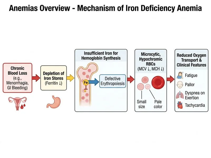
Sign up free to access AI-powered MCQ practice with detailed explanations and adaptive learning.