NEETPGAI
FeaturesNEET PGFMGEINI-CETBlogPricing
Log inStart Free
NEETPGAI

AI-powered NEET PG preparation platform. Master all 19 subjects with adaptive MCQs, AI tutoring, and spaced repetition.

Product

  • Features
  • Subjects
  • Previous Year Questions
  • NEET PG Preparation
  • FMGE Preparation
  • INI-CET Preparation
  • Compare
  • Pricing
  • Blog

Features

  • Adaptive MCQ Practice
  • AI Tutor
  • Mock Tests
  • Spaced Repetition

Resources

  • Blog
  • Study Guides
  • NEET PG Updates
  • Contact & support

Legal

  • Privacy Policy
  • Terms of Service

Stay updated

© 2026 NEETPGAI. All rights reserved.
    Subjects/Anatomy/Brachial Plexus
    Brachial Plexus
    hard
    bone Anatomy

    A 52-year-old man presents to the orthopedic clinic with a 3-month history of progressive wasting of the thenar and hypothenar eminences bilaterally. He reports a history of cervical rib on the left side, which was noted on chest X-ray. On examination, he has claw hand deformity affecting the 4th and 5th digits bilaterally, with sensory loss over the medial aspect of both forearms. He denies any neck trauma or birth injury. What is the anatomical basis for this bilateral presentation?

    A. Bilateral ulnar nerve compression at the cubital tunnel
    B. Bilateral C8 and T1 nerve root compression at the thoracic outlet
    C. Bilateral musculocutaneous nerve entrapment at the elbow
    D. Bilateral median nerve compression at the wrist

    Explanation

    Thoracic Outlet Syndrome (TOS) – Lower Trunk Compression

    This patient presents with neurogenic TOS affecting the lower trunk of the brachial plexus bilaterally.

    Clinical Features Explained

    Motor Findings:

    • Thenar and hypothenar wasting → Intrinsic hand muscles (C8, T1)
      • Thenar: Abductor pollicis brevis, flexor pollicis brevis, opponens pollicis (median and ulnar)
      • Hypothenar: Abductor digiti minimi, flexor digiti minimi, opponens digiti minimi (ulnar)
    • Claw hand (4th and 5th digits) → Interossei and lumbricals (C8, T1 via ulnar nerve)

    Sensory Findings:

    • Medial forearm sensory loss → Medial antebrachial cutaneous nerve (C8, T1)
    Anatomical Basis for Bilateral Presentation
    Loading diagram...
    Key Point
    Cervical rib is the most common anatomical cause of neurogenic TOS. It arises from the C7 transverse process and can compress the lower trunk (C8–T1) as it passes over the rib. Bilateral presentation suggests either bilateral cervical ribs or bilateral fibrous bands.
    High-YieldNEET PG
    Lower trunk (C8–T1) compression produces:
    1. 1.
      Intrinsic hand muscle atrophy (thenar + hypothenar)
    2. 2.
      Claw hand (especially 4th and 5th digits)
    3. 3.
      Medial forearm sensory loss (C8–T1 dermatome)
    4. 4.
      Horner's syndrome (if T1 root severely compressed)

    Mnemonic: LOAF — Lumbricals (C8–T1), Opponens pollicis (C8–T1), Abductor pollicis brevis (C8–T1), Flexor pollicis brevis (C8–T1) — all supplied by C8–T1 and affected in lower trunk injury.

    Clinical Pearl
    The bilateral nature is the key diagnostic clue—it points to a structural anatomical abnormality (cervical ribs, fibrous bands) rather than a single traumatic event. Unilateral TOS is more commonly from trauma or postural compression.
    Differential Diagnosis
    Table
    ConditionMotor LossSensory LossBilateral?Cause
    Lower trunk TOSIntrinsic hand muscles (C8–T1)Medial forearm (C8–T1)OftenCervical rib, fibrous band
    Ulnar nerve (cubital tunnel)Intrinsic hand musclesMedial 1.5 digitsRareCompression at elbow
    Median nerve (carpal tunnel)Thenar onlyLateral 3.5 digitsPossibleCompression at wrist
    Musculocutaneous entrapmentBiceps, brachialisLateral forearmRareCompression in arm

    Loading illustration…Brachial Plexus diagram

    Practice similar questions

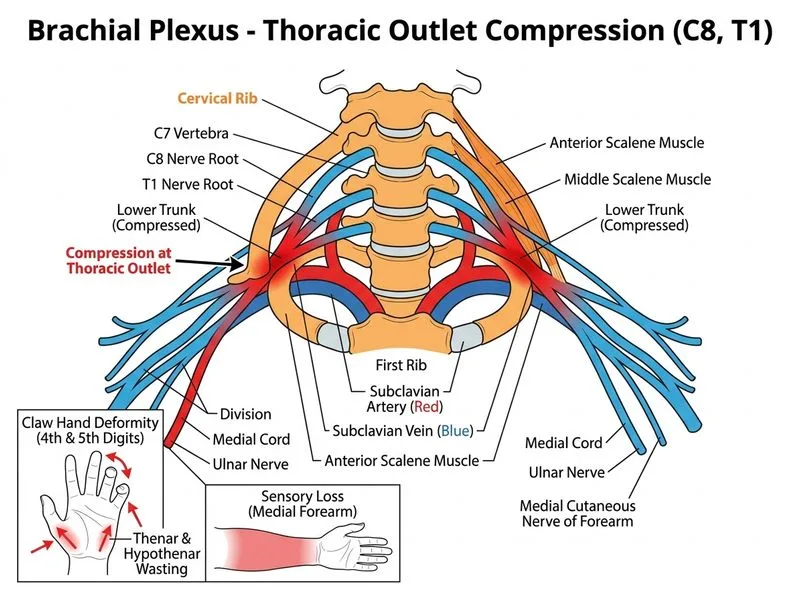
    Sign up free to access AI-powered MCQ practice with detailed explanations and adaptive learning.

    Start Practicing Free More Anatomy Questions

    Join our NEET PG community

    Daily MCQs, study tips, and topper strategies on Telegram.

    Join on Telegram →