Rheumatic mitral stenosis (MS) with new-onset atrial fibrillation and haemodynamic instability requires rapid, non-invasive assessment of valve area and transmitral gradients to guide urgent intervention (rate control, anticoagulation, or mechanical relief).
| Advantage | Relevance |
|---|---|
| Non-invasive | Safe in acute haemodynamic instability; no contrast or arterial access |
| Rapid | Can be performed at bedside; results available within 30 min |
| Quantifies severity | MVA and gradients guide urgency of intervention |
| Assesses complications | Detects LA thrombus, RV dysfunction, pulmonary hypertension |
| Guides intervention | Echo findings determine suitability for PMBV vs. surgery |
| Investigation | Role in Acute MS + AF |
|---|---|
| Transthoracic echo | Gold standard; rapid, quantifies severity, guides intervention |
| Cardiac catheterisation | Invasive; reserved for cases where echo is inconclusive or when coronary anatomy needed before surgery |
| Chest X-ray | Shows pulmonary congestion, LA enlargement; does NOT quantify MS severity |
| ECG | Shows AF, LAD; does NOT assess valve haemodynamics |
| Transoesophageal echo | Better for LA thrombus detection if PMBV planned; not first-line for severity assessment |
Loading illustration…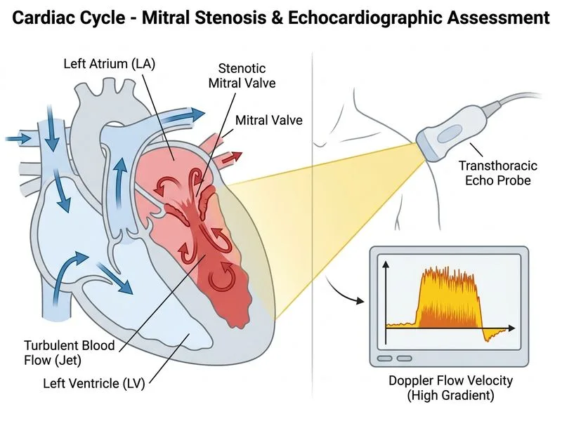
Sign up free to access AI-powered MCQ practice with detailed explanations and adaptive learning.
Daily MCQs, study tips, and topper strategies on Telegram.
Join on Telegram →