| Feature | Hyperplasia | Hypertrophy |
|---|---|---|
| Definition | ↑ Number of cells | ↑ Size of individual cells |
| Mechanism | Increased mitotic activity (controlled) | Increased protein synthesis per cell |
| Cell count | Elevated | Normal |
| Individual cell size | Normal | Enlarged |
| Reversibility | Reversible if stimulus removed | Reversible if stimulus removed |
| Example | Leiomyoma, endometrial hyperplasia | Cardiac hypertrophy, skeletal muscle |
The distinction matters: hyperplasia = benign proliferation with normal cell morphology, whereas dysplasia = abnormal morphology with increased mitotic rate and atypia (premalignant). This fibroid shows none of the latter features.
Uterine fibroids arise from:
Mnemonic: HyPERplasia = PERcentage of cells ↑
Loading illustration…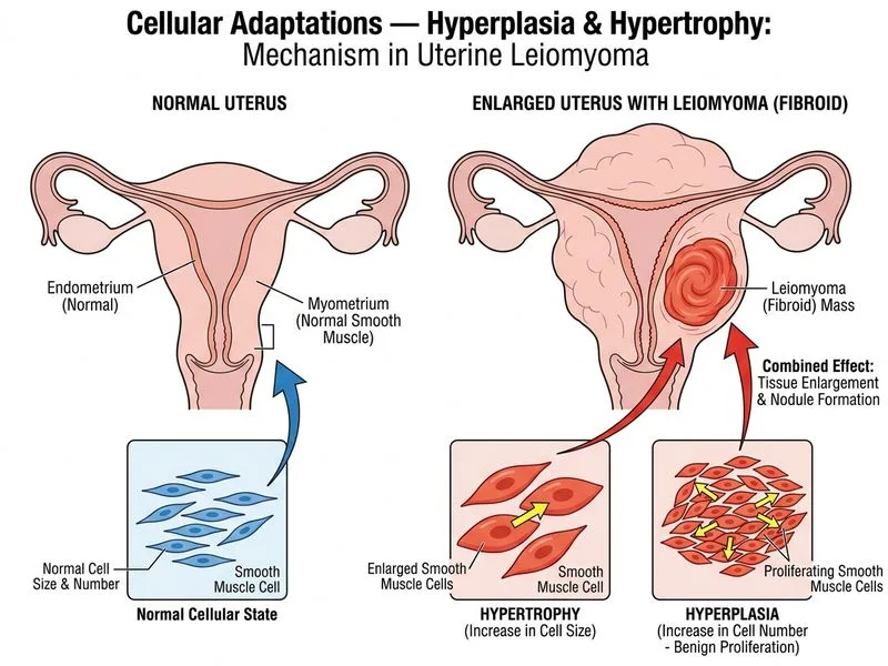
Sign up free to access AI-powered MCQ practice with detailed explanations and adaptive learning.
Daily MCQs, study tips, and topper strategies on Telegram.
Join on Telegram →