Chronic Suppurative Otitis Media MCQ — NEET PG Practice Question | NEETPGAI
Chronic Suppurative Otitis Media
hard
ear ENT
A 28-year-old woman with a 5-year history of chronic ear discharge and conductive hearing loss is being evaluated for possible cholesteatoma. Otoscopy shows a retraction pocket with foul-smelling discharge and granulation tissue. Which investigation is most specific for confirming the diagnosis of cholesteatoma?
A. Otoacoustic emissions and tympanometry
B. Diffusion-weighted MRI (DW-MRI) of the temporal bone
C. Conventional radiography of the mastoid (Schuller's view)
D. High-resolution CT (HRCT) of the temporal bone
Explanation
Imaging Modality for Cholesteatoma Confirmation
Key Point
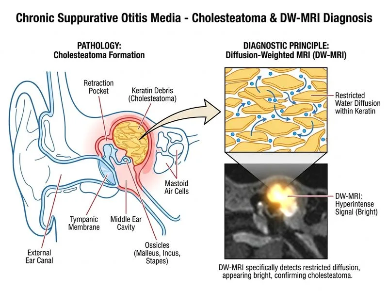
Diffusion-weighted MRI (DW-MRI) is the most specific and sensitive non-invasive investigation for confirming the presence of cholesteatoma, especially in cases where clinical suspicion is high but imaging findings are equivocal.
Why DW-MRI is Superior for Cholesteatoma
High-YieldNEET PG
DW-MRI exploits the fact that cholesteatoma contains desquamated keratin and cholesterol, which show restricted diffusion (high signal on DW sequences and low apparent diffusion coefficient [ADC]):
Sensitivity: 90–100% for detecting cholesteatoma
Specificity: 95–100% (can differentiate cholesteatoma from granulation tissue or other middle ear masses)
Non-invasive: No radiation exposure
Real-time assessment: Can detect both osseous and soft tissue disease
Clinical Pearl
In this patient with a retraction pocket and granulation tissue, DW-MRI will definitively confirm whether cholesteatoma is present. This is critical because cholesteatoma is a bone-eroding lesion that requires surgical removal, whereas simple chronic suppurative otitis media with granulation tissue may be managed medically or with less extensive surgery.
Comparison of Imaging Modalities
Table
Investigation
Sensitivity for Cholesteatoma
Specificity
Radiation
Best Use
DW-MRI
90–100%
95–100%
None
Confirmatory test; differentiates from granulation tissue
HRCT
70–85%
80–90%
Yes
Bone erosion, ossicular status, surgical planning
Conventional X-ray (Schuller's)
50–60%
60–70%
Yes
Mastoid sclerosis; poor sensitivity for soft tissue
Otoacoustic emissions
N/A
N/A
None
Cochlear function only; no role in cholesteatoma diagnosis
Mnemonic
DW-MRI = Definitive for Cholesteatoma (restricted diffusion = keratin/cholesterol = high specificity)
Diagnostic Algorithm
Loading diagram...
Why DW-MRI Over HRCT Alone
While HRCT is excellent for assessing bone erosion and ossicular involvement, it cannot reliably differentiate cholesteatoma from:
Granulation tissue
Cholesteatoma debris
Pus-filled retraction pockets
DW-MRI's restricted diffusion property makes it uniquely specific for cholesteatoma's keratinous content.
Loading illustration…
Practice similar questions
Sign up free to access AI-powered MCQ practice with detailed explanations and adaptive learning.