## Fracture-Related Infection (FRI) / Post-Traumatic Osteomyelitis — Pathogenesis and Diagnosis ### Clinical Context This patient has a **closed fracture** (no break in skin at time of injury) with signs of infection appearing **3 days post-injury**. The combination of: - **Fever and systemic signs** (malaise, elevated WBC 14,000/μL, elevated ESR/CRP) - **Local signs** (swelling, warmth over fracture site) - **Radiological evidence** (periosteal reaction, cortical lucencies) within days - **Closed fracture** (no open wound, no known bacteremia source) ...points to **Fracture-Related Infection (FRI)** — also termed **post-traumatic osteomyelitis** in traditional Indian curricula (Maheshwari's Essential Orthopaedics) — rather than hematogenous osteomyelitis or cellulitis. ### Why NOT the Other Options? | Option | Reason Excluded | |--------|----------------| | **A — Hematogenous osteomyelitis** | Requires a distant bacteremia source (e.g., skin infection, UTI, endocarditis) or immunosuppression — none mentioned. Typically affects metaphysis of long bones in children without preceding fracture. | | **B — Fat embolism syndrome** | Presents 24–72 h post-fracture with **respiratory distress, petechiae, and confusion** (Gurd's criteria) — not with localized bone infection signs or radiological lucencies. | | **D — Cellulitis** | Soft-tissue infection only; does **not** produce periosteal reaction or cortical lucencies on plain radiograph. Systemic markers are usually milder, and there is no bone involvement. | ### Pathogenesis of FRI in Closed Fractures **Key Point:** Even in closed fractures, bacteria can be introduced at the moment of injury via **contaminated bone fragments, soil, or soft-tissue trauma** (micro-contamination). The avascular, hypoxic fracture hematoma provides an ideal niche for bacterial proliferation and biofilm formation, shielded from host defenses. - **Route:** Local contamination at fracture site (not hematogenous) - **Timing:** Days to weeks post-injury (early FRI: < 2 weeks) - **Common organisms:** *S. aureus* (most common), gram-negatives, anaerobes - **Radiological onset:** Periosteal reaction and cortical lucencies as early as **3–7 days** (earlier than hematogenous osteomyelitis, which typically shows changes at 10–14 days) ### Diagnostic Confirmation 1. **Blood cultures** — may be positive if bacteremia has developed 2. **Fracture aspirate / surgical specimen culture** — gold standard 3. **Imaging:** - **Plain radiographs:** Periosteal reaction, cortical lucencies, widened fracture line (as seen here) - **MRI:** Best for early marrow edema and soft-tissue extent - **CT:** Useful for sequestrum/involucrum in established cases 4. **Labs:** Elevated WBC, ESR, CRP — non-specific but supportive; serial CRP most useful for monitoring **Clinical Pearl (Harrison's / Maheshwari):** Early recognition and **aggressive surgical debridement** (removal of devitalized bone and soft tissue) combined with **prolonged antibiotics** (4–6 weeks IV, then oral) are essential to prevent progression to **chronic osteomyelitis** and **non-union** — the two most feared complications. ### Management Summary 1. Blood cultures + fracture aspirate before antibiotics 2. Empiric broad-spectrum IV antibiotics (e.g., cloxacillin + gentamicin), then tailor to culture sensitivities 3. Surgical debridement of infected/devitalized bone and soft tissue 4. Fracture stabilization (ORIF or external fixation) 5. Monitor with serial CRP, ESR, and imaging **High-Yield for NEET PG:** In a closed fracture with early fever, local bone signs, and radiological changes — the answer is **FRI / post-traumatic osteomyelitis (Option C)**. Hematogenous osteomyelitis requires a bacteremia source; cellulitis lacks bone radiological changes.
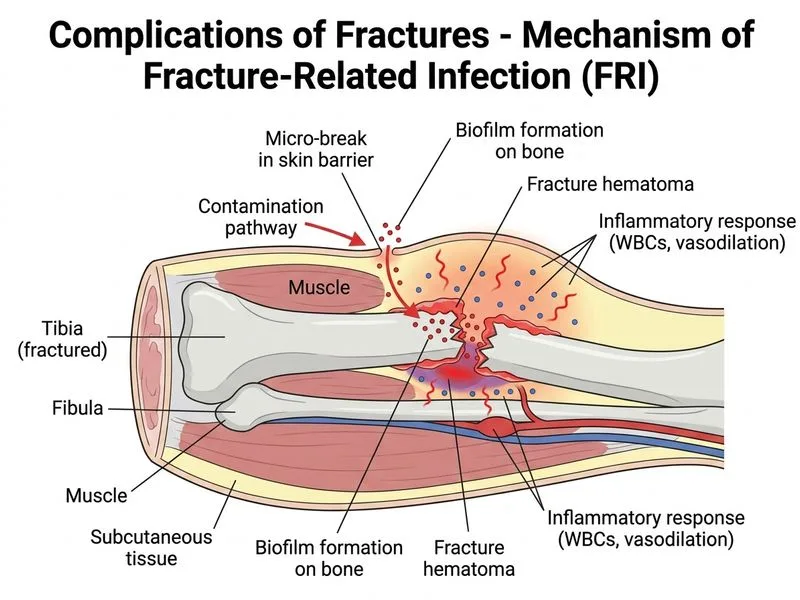
Sign up free to access AI-powered MCQ practice with detailed explanations and adaptive learning.