Which of the following radiological signs is pathognomonic for pulmonary collapse rather than consolidation on chest X-ray?
A. Silhouette sign
B. Opacity obscuring the hilum
C. Air bronchogram
D. Loss of volume with displacement of fissures
Explanation
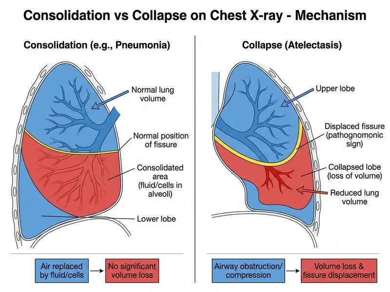
Distinguishing Collapse from Consolidation
Key Point
The hallmark of collapse is loss of volume with secondary displacement of anatomical structures (fissures, mediastinum, diaphragm), whereas consolidation maintains normal or increased volume.
Radiological Signs Comparison
Table
Feature
Consolidation
Collapse
Volume
Normal or increased
Decreased
Fissures
Normal position
Displaced toward lesion
Air bronchogram
Present (pathognomonic)
Absent or rare
Silhouette sign
May occur
May occur
Mediastinal shift
None
Toward affected side
Diaphragm
Normal position
Elevated on affected side
High-YieldNEET PG
Loss of volume with fissural displacement is the ONLY sign that definitively indicates collapse and excludes consolidation. Consolidation can mimic collapse in appearance, but volume loss is the discriminator.
Why This Matters Clinically
Clinical Pearl
Consolidation suggests infection, aspiration, or infarction (parenchymal process). Collapse suggests airway obstruction (tumor, mucus plug, foreign body) requiring different management.
Mnemonic — COLLAPSE signs:
Contraction (volume loss)
Obliteration of fissures (displacement)
Loss of normal landmarks
Lateral shift of mediastinum
Apex elevation (in upper lobe)
Posterior displacement of fissures
Silhouette may occur
Elevation of hemidiaphragm
Felson's Principles of Chest Roentgenology
Loading illustration…
Practice similar questions
Sign up free to access AI-powered MCQ practice with detailed explanations and adaptive learning.