Consolidation vs Collapse on Chest X-ray MCQ — NEET PG Practice Question | NEETPGAI
Consolidation vs Collapse on Chest X-ray
medium
scan Radiology
A 62-year-old man with COPD and a 40-pack-year smoking history presents with acute onset dyspnea and cough productive of purulent sputum. On examination, he is febrile (38.5°C), with decreased breath sounds and dullness to percussion over the right lower lobe. Chest X-ray shows an area of increased opacity in the right lower lobe with air bronchograms visible within it. The hilum and mediastinum are in their normal positions. What is the most likely radiological finding?
A. Right lower lobe pleural effusion
B. Right lower lobe consolidation
C. Right lower lobe collapse
D. Right lower lobe pneumothorax
Explanation
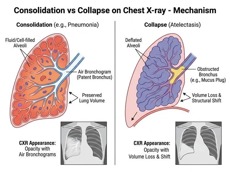
Consolidation vs Collapse: Key Radiological Distinction
Key Point
The presence of air bronchograms within an opacified area is pathognomonic for consolidation, not collapse. Air bronchograms represent air-filled bronchi silhouetted against consolidated (fluid-filled) alveoli.
Radiological Features of Consolidation
Table
Feature
Consolidation
Collapse
Air bronchograms
Present (hallmark)
Absent
Hilum position
Normal
Shifted toward affected lobe
Mediastinum
Normal
May shift toward affected side
Heart border
May be obscured (silhouette sign)
Usually clear
Density
Homogeneous opacity
Wedge-shaped or linear opacity
Diaphragm
Normal position
Elevated on affected side
High-YieldNEET PG
Air bronchograms are the single most reliable sign distinguishing consolidation from other opacities. They indicate patent airways within consolidated lung parenchyma.
Clinical Correlation
This patient has:
Fever (38.5°C) — suggests infection
Purulent sputum — indicates bacterial pneumonia
Dullness to percussion — consistent with fluid-filled (consolidated) lung
Decreased breath sounds — from alveolar filling
Normal hilum and mediastinum — rules out collapse (which causes mediastinal shift)
Clinical Pearl
In consolidation, the volume of the affected lobe remains normal because alveoli are filled with inflammatory exudate, not collapsed. In collapse, alveolar air is reabsorbed, causing volume loss and characteristic mediastinal/hilar shift.
Mnemonic: ABC of Consolidation
Air bronchograms present
Bronchi patent and visible
Complete alveolar filling with fluid/pus
Why This Is Consolidation and Not Collapse
1.
Air bronchograms visible — consolidation hallmark
2.
Normal hilum position — collapse causes hilar shift
3.
Normal mediastinum — collapse causes mediastinal shift
4.
Clinical presentation — acute pneumonia with fever and purulent sputum
Felson's Principles of Chest Roentgenology Ch 2
Loading illustration…
Practice similar questions
Sign up free to access AI-powered MCQ practice with detailed explanations and adaptive learning.