Consolidation vs Collapse on Chest X-ray MCQ — NEET PG Practice Question | NEETPGAI
Consolidation vs Collapse on Chest X-ray
hard
scan Radiology
A 58-year-old woman with advanced lung cancer presents with progressive dyspnea over 3 days. On examination, she has reduced breath sounds on the left side with dullness to percussion. Chest X-ray reveals a wedge-shaped opacity in the left lower lobe with the apex pointing toward the hilum. The left hilum is displaced medially, and the left hemidiaphragm is elevated. The mediastinum is shifted toward the left. No air bronchograms are visible within the opacity. What is the most likely radiological diagnosis?
A. Left lower lobe collapse due to endobronchial obstruction
B. Left lower lobe consolidation due to post-obstructive pneumonia
C. Left lower lobe pneumonia with septal thickening
D. Left lower lobe pleural effusion with atelectasis
Explanation
Collapse: Radiological Signs and Mechanisms
Key Point
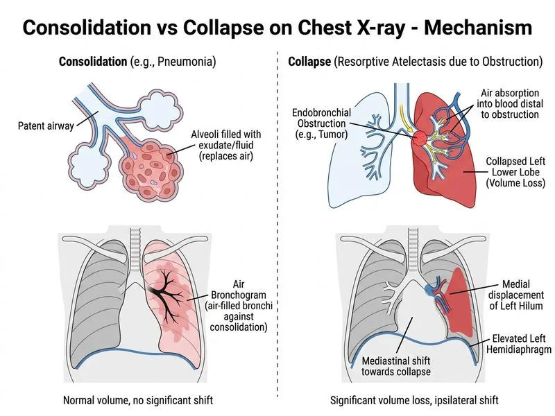
Collapse (atelectasis) is characterized by volume loss with mediastinal/hilar shift, elevated hemidiaphragm, and absence of air bronchograms. The wedge-shaped opacity with apex toward the hilum is classic for lobar collapse.
Radiological Features of Collapse
Table
Feature
Collapse
Consolidation
Shape
Wedge-shaped, apex toward hilum
Homogeneous, amorphous
Hilar shift
Toward affected lobe (medial in left lower)
No shift
Mediastinal shift
Toward affected side
No shift
Hemidiaphragm
Elevated on affected side
Normal position
Air bronchograms
Absent
Present
Volume
Decreased (loss of air)
Normal
High-YieldNEET PG
The combination of mediastinal shift + hilar shift + elevated hemidiaphragm + absent air bronchograms = collapse, not consolidation.
Pathophysiology of Collapse in Lung Cancer
1.
Endobronchial obstruction — tumor occludes left lower lobe bronchus
2.
Air absorption — distal air is reabsorbed into bloodstream
Hilar displacement — hilum moves medially with collapsing lobe
Clinical Pearl
In malignancy, collapse is often due to endobronchial tumor obstruction. Post-obstructive pneumonia may develop distal to the obstruction, but the primary finding here is collapse (wedge shape, shift, no air bronchograms).