NEETPGAI
FeaturesNEET PGFMGEINI-CETBlogPricing
Log inStart Free
NEETPGAI

AI-powered NEET PG preparation platform. Master all 19 subjects with adaptive MCQs, AI tutoring, and spaced repetition.

Product

  • Features
  • Subjects
  • Previous Year Questions
  • NEET PG Preparation
  • FMGE Preparation
  • INI-CET Preparation
  • Compare
  • Pricing
  • Blog

Features

  • Adaptive MCQ Practice
  • AI Tutor
  • Mock Tests
  • Spaced Repetition

Resources

  • Blog
  • Study Guides
  • NEET PG Updates
  • Contact & support

Legal

  • Privacy Policy
  • Terms of Service

Stay updated

© 2026 NEETPGAI. All rights reserved.
    Subjects/Radiology/Consolidation vs Collapse on Chest X-ray
    Consolidation vs Collapse on Chest X-ray
    medium
    scan Radiology

    A 58-year-old woman with lung cancer undergoes right upper lobe resection. Post-operative day 3 chest X-ray shows opacity in the right hemithorax with the right heart border remaining visible and the right hemidiaphragm in normal position. Which investigation is most appropriate to determine whether this represents post-operative consolidation or atelectasis?

    A. CT chest with IV contrast
    B. Bronchoscopy with bronchoalveolar lavage
    C. Ventilation-perfusion scan
    D. Lateral decubitus chest X-ray

    Explanation

    Post-operative Opacity: Consolidation vs. Atelectasis

    Key Point
    In the post-operative setting, distinguishing consolidation from atelectasis is critical for management. CT chest with IV contrast is the investigation of choice because it can definitively characterize the opacity and guide clinical decision-making.
    Clinical Context: Post-operative Day 3

    After lung resection, opacities can represent:

    • Atelectasis (most common, especially in early post-op period)
    • Consolidation (aspiration, pneumonia, pulmonary edema)
    • Pleural effusion
    • Hemorrhage
    High-YieldNEET PG
    The absence of silhouette sign (right heart border visible) and normal hemidiaphragm position make simple atelectasis less likely, but this does not exclude it entirely. CT is needed for definitive characterization.
    Why CT Chest with IV Contrast is Superior
    Table
    FeatureCT Advantage
    Tissue characterizationDistinguishes consolidation (homogeneous, air bronchograms) from atelectasis (wedge-shaped, no air bronchograms)
    Contrast enhancementConsolidation enhances; atelectasis does not (or minimally)
    Vascular involvementDetects pulmonary embolism (post-op risk)
    Pleural/mediastinal assessmentEvaluates for effusion, hematoma, or mediastinal shift
    Exclusion of complicationsPneumonia, abscess, bronchopleural fistula
    Clinical Pearl
    In post-operative patients, IV contrast CT can simultaneously assess for PE (a major post-operative complication) while characterizing the lung opacity—making it the most efficient and comprehensive choice.
    Why Other Investigations Are Inadequate

    Lateral decubitus X-ray: Useful only for detecting free pleural fluid; does not characterize parenchymal consolidation or atelectasis in the post-operative setting.

    Bronchoscopy with BAL: Invasive and non-specific; used therapeutically (to clear secretions in atelectasis) or diagnostically (to obtain cultures), but does not definitively characterize the opacity on imaging.

    Ventilation-perfusion scan: Outdated for this purpose; CT pulmonary angiography (CTPA) has replaced V/Q scanning for PE detection, and CT chest is superior for parenchymal characterization.

    Mnemonic: CT for Post-op Opacity — Characterization, Tissue assessment, Exclusion of PE and complications.

    Loading illustration…Consolidation vs Collapse on Chest X-ray diagram

    Practice similar questions

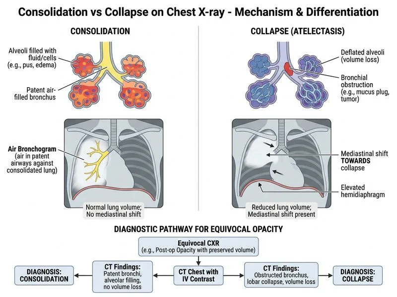
    Sign up free to access AI-powered MCQ practice with detailed explanations and adaptive learning.

    Start Practicing Free More Radiology Questions

    Join our NEET PG community

    Daily MCQs, study tips, and topper strategies on Telegram.

    Join on Telegram →