Air bronchogram represents air-filled bronchi visualized against a background of consolidated (fluid-filled) lung parenchyma. This finding is pathognomonic for consolidation and occurs because:
In atelectasis (collapse), air bronchograms are typically absent because:
| Feature | Consolidation | Atelectasis |
|---|---|---|
| Air bronchogram | Present ✓ | Absent |
| Mediastinal shift | Toward normal side (away) | Toward affected side (toward) |
| Volume of affected lobe | Normal or increased | Decreased |
| Silhouette sign | May be present | May be present |
| Bronchial breath sounds | Present | May be present |
| Tactile fremitus | Increased | Decreased |
Loading illustration…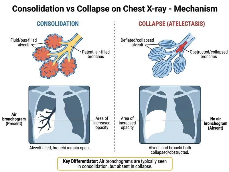
Sign up free to access AI-powered MCQ practice with detailed explanations and adaptive learning.
Daily MCQs, study tips, and topper strategies on Telegram.
Join on Telegram →