Tinea pedis caused by Trichophyton rubrum typically presents with which clinical pattern?
A. Annular plaques with central clearing on the sole
B. Acute vesiculobullous eruption on the dorsum of the foot
C. Kerion-like pustular lesion with lymphadenopathy
D. Chronic interdigital maceration with scaling and fissuring
Explanation
Clinical Presentation of Tinea Pedis by Organism
Key Point
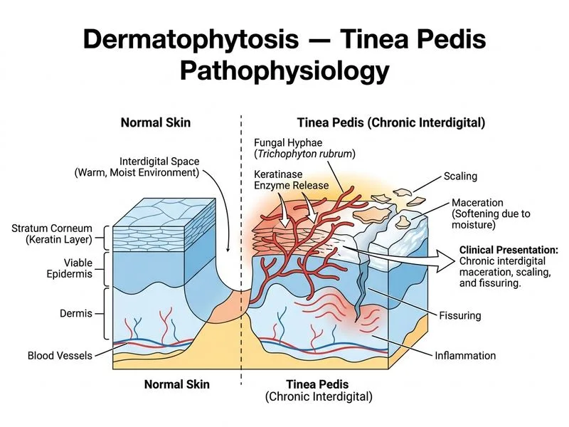
The clinical morphology of tinea pedis varies with the causative organism. T. rubrum causes a chronic, indolent infection characterized by interdigital involvement with minimal inflammation.
High-YieldNEET PG
T. rubrum tinea pedis is the most common type worldwide and typically presents as a chronic interdigital dermatophytosis with maceration, scaling, and fissuring, often affecting the fourth and fifth toe webs first.
Three Main Clinical Patterns of Tinea Pedis
Table
Pattern
Organism
Presentation
Inflammation
Duration
Interdigital (most common)
T. rubrum, T. mentagrophytes var. mentagrophytes
Maceration, scaling, fissuring in toe webs (4th–5th web first)
Minimal to mild
Chronic, indolent
Plantar/Moccasin type
T. rubrum
Diffuse scaling on sole and lateral foot; "moccasin" distribution
Minimal
Chronic, very resistant to treatment
Vesiculobullous/Acute
T. mentagrophytes var. mentagrophytes
Vesicles and bullae, often on arch or lateral sole
Marked
Acute, often secondary bacterial infection
Clinical Pearl
T. rubrum causes a dry, scaly interdigital infection, whereas T. mentagrophytes causes an acute, inflammatory vesiculobullous pattern. The difference reflects their anthropophilic vs. zoophilic adaptation.
Mnemonic — "TRIM" for T. rubrum Tinea Pedis:
T — Toe webs (interdigital)
R — Rubrum (T. rubrum)
I — Indolent (chronic, minimal inflammation)
M — Maceration and scaling
Why Interdigital Involvement?
1.
Toe webs provide warm, moist, occluded environment
2.
Fourth and fifth toe webs are most occluded
3.
T. rubrum thrives in chronic, low-inflammation conditions