A 28-year-old male farmer from rural Maharashtra presents with a 3-week history of itchy, scaly patches on his right groin and inner thigh. The lesions are well-demarcated, erythematous, with a raised border and central clearing. He denies systemic symptoms. KOH mount of scale shows septate hyphae. Which of the following is the most likely causative organism?
A. Microsporum canis
B. Candida albicans
C. Epidermophyton floccosum
D. Trichophyton mentagrophytes
Explanation
Diagnosis: Tinea Cruris (Jock Itch)
Clinical Presentation
The patient presents with classic features of tinea cruris:
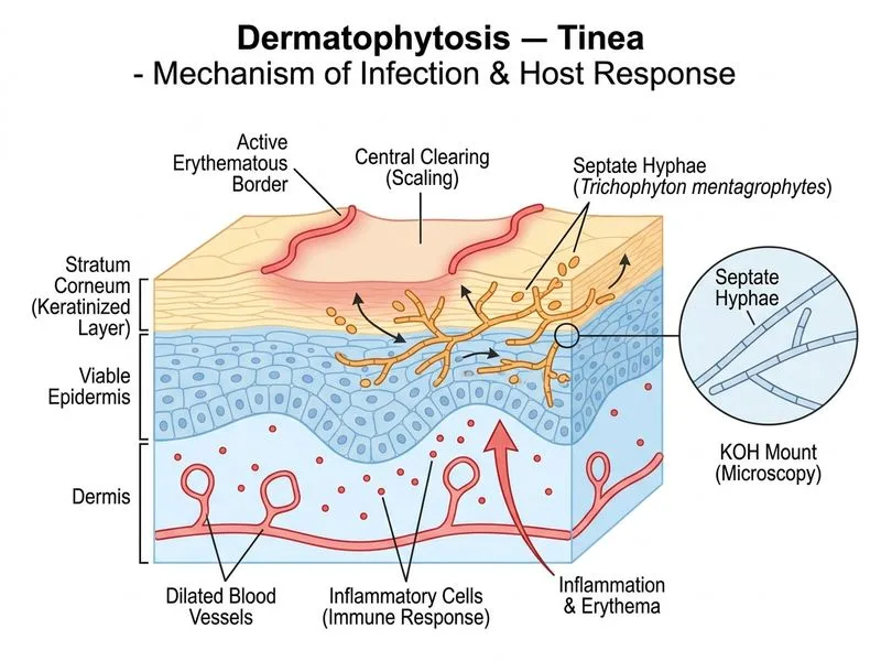
Well-demarcated erythematous patches with raised borders and central clearing ("ringworm" appearance)
Intense pruritus localized to groin and inner thigh (warm, moist intertriginous area)
Risk factors: male sex, warm climate, occupational exposure (farmer)
KOH mount showing septate hyphae confirms a dermatophyte (rules out Candida)
Causative Organisms in Tinea Cruris
Table
Organism
Frequency (Global/India)
Notes
Trichophyton rubrum
Most common overall (~50–60%)
Anthropophilic; most frequent cause worldwide
Epidermophyton floccosum
Second most common (~20–30%)
Exclusively causes tinea cruris & tinea pedis; no hair invasion
Trichophyton mentagrophytes
Less common in cruris
More common in tinea pedis/unguium
Microsporum canis
Rare in groin
Zoophilic; primarily scalp/body
Key Point
Epidermophyton floccosum is classically highlighted as the second most common cause of tinea cruris and is the organism most specifically associated with the groin in standard dermatology and mycology textbooks (Fitzpatrick's Dermatology, Rippon's Medical Mycology). Among the options provided — where T. rubrum is absent — E. floccosum is the best answer, as T. mentagrophytes is primarily associated with tinea pedis and tinea unguium rather than tinea cruris.
Why NOT the other options?
Trichophyton mentagrophytes (A): More commonly causes tinea pedis ("athlete's foot") and onychomycosis; less frequently implicated in tinea cruris compared to E. floccosum.
Microsporum canis (B): Zoophilic dermatophyte; primarily causes tinea capitis and tinea corporis; rarely causes tinea cruris.
Candida albicans (D): Produces pseudohyphae and budding yeast on KOH — not septate hyphae. Candidal intertrigo lacks the classic raised, scaly border with central clearing.
KOH Mount Findings
High-YieldNEET PG
Septate hyphae on KOH preparation confirm a dermatophyte infection. Epidermophyton floccosum characteristically produces club-shaped macroconidia in clusters (2–4) and no microconidia — a distinguishing feature in culture.
Pathophysiology
1.
Dermatophyte colonizes stratum corneum via keratinolytic enzymes
2.
Triggers inflammatory response → erythema and scaling
3.
Central clearing due to host T-cell–mediated immunity
Clinical Pearl
Epidermophyton floccosum is unique among dermatophytes in that it does not infect hair — it is restricted to skin and nails. This makes it a classic cause of tinea cruris and tinea pedis. (Reference: Fitzpatrick's Dermatology, 9th ed.; Rippon JW, Medical Mycology, 3rd ed.)