| Feature | Tinea Corporis | Tinea Cruris |
|---|---|---|
| Location | Trunk, limbs, face | Inguinal folds, inner thighs, perianal |
| Morphology | Annular with central clearing | Sharply demarcated, often bilateral |
| Border | Raised, erythematous, scaly | Sharp, well-defined, may show maceration |
| Maceration | Absent | Present (due to moisture & friction) |
| Pruritus | Mild to moderate | Severe |
Both conditions can present with annular lesions and central clearing. This is a common morphology in dermatophytosis but does not distinguish between the two sites.
While these are risk factors for both conditions, they are not discriminating features—both tinea corporis and tinea cruris are predisposed by these factors.
Loading illustration…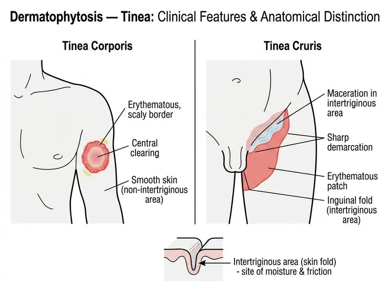
Sign up free to access AI-powered MCQ practice with detailed explanations and adaptive learning.
Daily MCQs, study tips, and topper strategies on Telegram.
Join on Telegram →