NEETPGAI
FeaturesNEET PGFMGEINI-CETBlogPricing
Log inStart Free
NEETPGAI

AI-powered NEET PG preparation platform. Master all 19 subjects with adaptive MCQs, AI tutoring, and spaced repetition.

Product

  • Features
  • Subjects
  • Previous Year Questions
  • NEET PG Preparation
  • FMGE Preparation
  • INI-CET Preparation
  • Compare
  • Pricing
  • Blog

Features

  • Adaptive MCQ Practice
  • AI Tutor
  • Mock Tests
  • Spaced Repetition

Resources

  • Blog
  • Study Guides
  • NEET PG Updates
  • Contact & support

Legal

  • Privacy Policy
  • Terms of Service

Stay updated

© 2026 NEETPGAI. All rights reserved.
    Subjects/Pathology/DIC
    DIC
    hard
    microscope Pathology

    A 52-year-old man with acute promyelocytic leukemia (APL) develops DIC after chemotherapy initiation. Which coagulation abnormality most reliably distinguishes APL-associated DIC from DIC secondary to sepsis?

    A. Thrombocytopenia with normal fibrinogen and low D-dimer
    B. Isolated prolongation of PT with normal aPTT and normal platelet count
    C. Severe hypofibrinogenemia with markedly elevated D-dimer and FDP
    D. Elevated tissue factor (TF) and cancer procoagulant (CP) with normal or mildly reduced factor V

    Explanation

    APL-Associated DIC vs. Sepsis-Associated DIC

    Pathophysiologic Basis
    Key Point
    APL-associated DIC is characterized by a hyperfibrinolytic phenotype driven by the release of procoagulant substances (tissue factor, cancer procoagulant) AND fibrinolytic activators (annexin II, tPA) from leukemic promyelocytes. This results in severe hypofibrinogenemia with markedly elevated D-dimer and FDP — the most reliable laboratory hallmark distinguishing APL-DIC from sepsis-DIC.
    Mechanism Comparison
    Table
    FeatureAPL-DICSepsis-DIC
    Primary driverTF + Cancer Procoagulant + Annexin II (fibrinolysis)Endotoxin → monocyte/endothelial TF expression
    Fibrinolysis↑↑↑ (hyperfibrinolytic)Variable (often suppressed by PAI-1)
    Fibrinogen↓↓↓ (severely low, often <100 mg/dL)↓↓ (moderate reduction)
    D-dimer / FDP↑↑↑ (markedly elevated)↑↑ (elevated, but less extreme)
    Platelets↓↓↓↓↓↓
    PT / aPTTProlongedProlonged
    Factor VConsumed (not reliably spared)Consumed
    Why Option A Is Correct
    High-YieldNEET PG
    The hallmark of APL-DIC is severe hypofibrinogenemia with markedly elevated D-dimer and FDP, reflecting the dominant hyperfibrinolytic component. APL promyelocytes release:
    1. 1.
      Annexin II — overexpressed on APL cell surfaces, acting as a co-receptor for plasminogen and tPA, dramatically accelerating plasmin generation and fibrinolysis
    2. 2.
      Tissue factor (TF) and cancer procoagulant (CP) — driving thrombin generation and fibrin formation, which is then rapidly lysed

    This combination of intense fibrin generation AND accelerated fibrinolysis produces profoundly low fibrinogen (often <50–100 mg/dL) and extremely high D-dimer/FDP — a degree of hypofibrinogenemia that is characteristically more severe than in typical sepsis-DIC, where PAI-1 elevation often suppresses fibrinolysis.

    Why Option B is incorrect: While APL cells do express elevated TF and cancer procoagulant, the claim that Factor V is "normal or mildly reduced" in APL-DIC is not supported by standard references (Williams Hematology, Harrison's). Factor V is consumed in both APL-DIC and sepsis-DIC; Factor V preservation is not a reliable distinguishing feature in clinical practice.

    Clinical Pearl
    APL-DIC is treated with ATRA + arsenic trioxide, which differentiate APL cells, reducing release of procoagulant and fibrinolytic mediators. Cryoprecipitate and fresh frozen plasma are used to replace fibrinogen and clotting factors. Antifibrinolytics (e.g., tranexamic acid) are used cautiously given the thrombotic risk.

    Reference: Levi M, Scully M. How I treat disseminated intravascular coagulation. Blood. 2018; Tallman MS, Altman JK. How I treat acute promyelocytic leukemia. Blood. 2009; Harrison's Principles of Internal Medicine, 21st ed., Chapter on Coagulation Disorders.

    Loading illustration…DIC diagram

    Practice similar questions

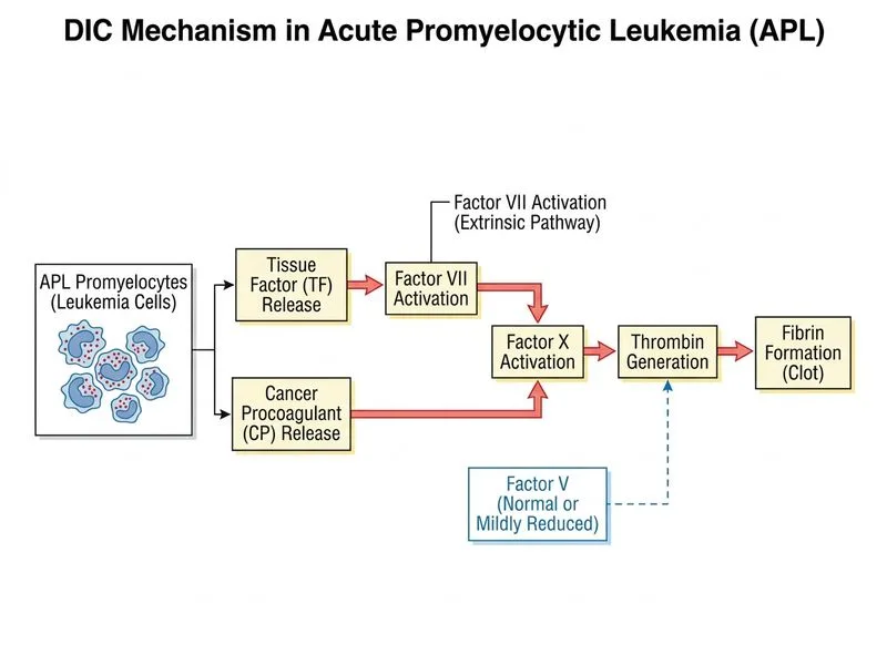
    Sign up free to access AI-powered MCQ practice with detailed explanations and adaptive learning.

    Start Practicing Free More Pathology Questions

    Join our NEET PG community

    Daily MCQs, study tips, and topper strategies on Telegram.

    Join on Telegram →