## Distinguishing AMI from LVH on ECG ### Key Diagnostic Features **Key Point:** ST-segment elevation with reciprocal ST-segment depression is pathognomonic for acute transmural myocardial infarction and is NOT seen in LVH, which is a chronic structural change. ### Comparison Table | Feature | Acute MI | LVH | | --- | --- | --- | | **ST-segment elevation** | Present (acute transmural injury) | Absent | | **Reciprocal ST depression** | Yes (in non-infarcted territory) | No | | **QRS voltage** | May be normal or low | Increased (Sokolow-Lyon, Cornell criteria) | | **T-wave changes** | Symmetrical inversion (acute phase) | Asymmetrical inversion (strain pattern) | | **Onset** | Acute (hours) | Gradual (months to years) | | **Evolutionary pattern** | Dynamic (ST→T changes over days) | Static | ### Why ST-Elevation is the Best Discriminator **High-Yield:** ST-segment elevation indicates acute transmural myocardial injury (necrosis of the full thickness of myocardium). This is an acute, dynamic process that does NOT occur in LVH, which is purely a chronic hypertrophic response to pressure overload. **Clinical Pearl:** The presence of reciprocal ST-segment depression (in leads opposite the infarct territory) further strengthens the diagnosis of AMI and is virtually never seen in LVH alone. ### Why Other Findings Are Shared or Non-Specific - **Increased QRS voltage:** Both AMI and LVH can show increased voltage, especially if the infarct is small or if LVH is severe. This is NOT discriminatory. - **T-wave inversion:** Can occur in both conditions. In AMI, it is symmetrical and evolves dynamically; in LVH, it is asymmetrical (strain pattern) and static. However, T-wave inversion alone does not reliably distinguish them. - **Left axis deviation:** More common in LVH but can occur in inferior MI or other conditions. Not specific enough to distinguish AMI from LVH. [cite:Harrison 21e Ch 297] 
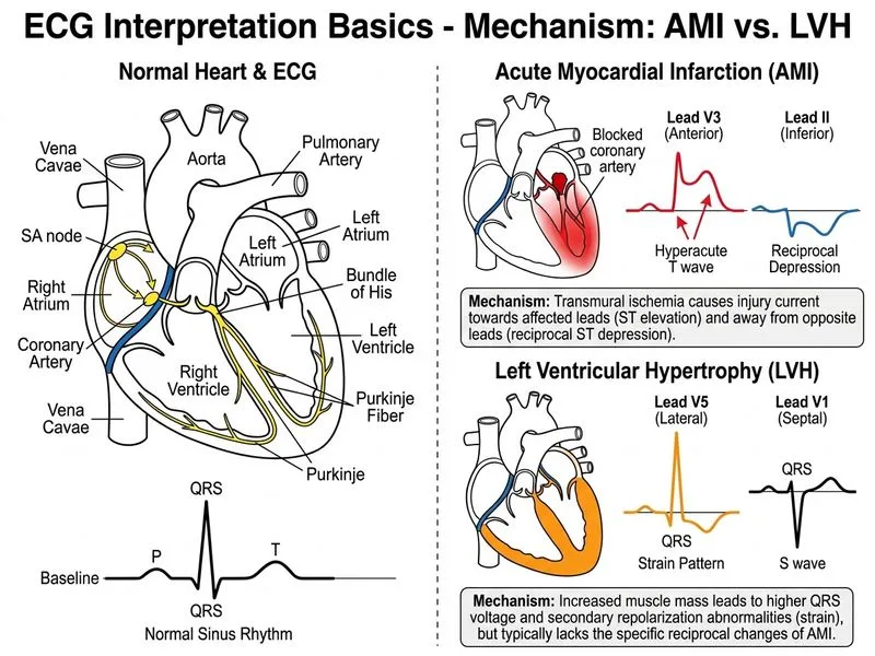
Sign up free to access AI-powered MCQ practice with detailed explanations and adaptive learning.