## Embryological Origin of Anterior Mediastinal Cyst The histological findings—stratified squamous epithelium, respiratory epithelium, smooth muscle, and cartilage—are pathognomonic for a **bronchogenic cyst**, which arises from abnormal budding of the **ventral foregut endoderm** during weeks 3–7 of gestation. ### Embryological Mechanism - The **primary bronchus** develops as a ventral outgrowth (diverticulum) from the foregut endoderm at the level of the pharynx (around week 4). - Abnormal or ectopic budding of this ventral diverticulum before complete separation of the respiratory diverticulum from the esophagus can result in bronchogenic cysts. - These cysts are lined with respiratory epithelium (ciliated columnar epithelium) and contain cartilage rings and smooth muscle—all derivatives of the foregut endoderm and its associated splanchnic mesoderm. ### Location & Clinical Significance - **Anterior mediastinal cysts** are the most common site (60–70% of bronchogenic cysts). - The cyst can compress the trachea, esophagus, or great vessels, causing respiratory distress or dysphagia. - Histology confirms the diagnosis: respiratory epithelium + cartilage + smooth muscle. ### Key Point **Bronchogenic cysts = ectopic foregut endoderm (specifically, abnormal budding of the respiratory diverticulum)** ### Mnemonic **"Broncho = Foregut Bud"** — Remember that the entire respiratory tree (larynx, trachea, bronchi, lungs) originates from the ventral foregut endoderm. 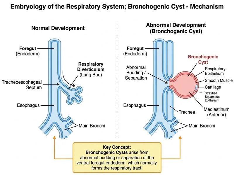
Sign up free to access AI-powered MCQ practice with detailed explanations and adaptive learning.