## Fracture Healing — Phases Overview **Key Point:** Fracture healing progresses through four distinct phases, each with characteristic cellular and tissue changes. ### Phase-by-Phase Timeline | Phase | Timing | Key Features | Callus Type | |-------|--------|--------------|-------------| | **Inflammatory** | 0–3 days | Hematoma formation, inflammatory cell infiltration, necrotic tissue removal | None yet | | **Soft Callus (Reparative)** | 1–3 weeks | Fibroblasts and chondrocytes proliferate; fibrocartilaginous matrix laid down; angiogenesis | Fibrocartilaginous | | **Hard Callus (Bridging)** | 3–12 weeks | Endochondral ossification; woven bone replaces cartilage; fracture site becomes rigid | Bony | | **Remodeling** | Months to years | Woven bone → lamellar bone; restoration of medullary canal; return to normal architecture | Mature bone | **High-Yield:** The **soft callus phase (1–3 weeks)** is when fibrocartilaginous callus forms—this is the bridge between inflammation and hard bone formation. The callus is still pliable and not yet rigid. **Clinical Pearl:** On X-ray, soft callus is often not visible because it is radiolucent (not calcified). Hard callus becomes radiographically apparent as ossification begins around week 3–4. **Mnemonic:** **I-S-H-R** = Inflammatory → Soft callus → Hard callus → Remodeling. Think: "Soft comes before Hard." ### Why This Phase Matters During the soft callus phase: - Fracture site is still mobile and vulnerable to re-injury. - Callus is composed mainly of fibrocartilage, not bone. - Endochondral ossification has not yet begun in significant amounts. - External callus is visible clinically (swelling) but not yet on plain radiographs. 
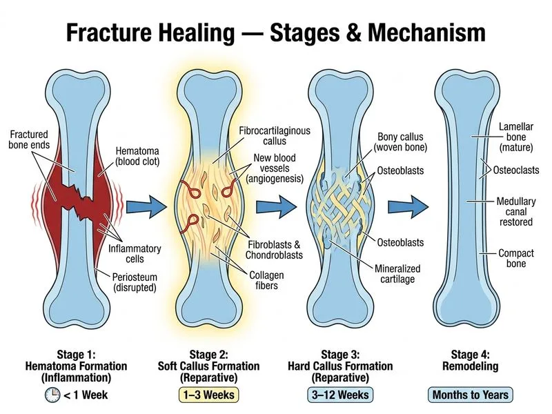
Sign up free to access AI-powered MCQ practice with detailed explanations and adaptive learning.