A 35-year-old male presents with a closed fracture of the mid-shaft humerus sustained 6 weeks ago. Clinical examination shows good callus formation and minimal tenderness. The patient is keen to know the stage of fracture healing and the best investigation to confirm the presence of bridging callus and assess fracture union.
A. Plain radiography (anteroposterior and lateral views)
B. MRI of the humerus
C. Ultrasound of the fracture site
D. CT scan with 3D reconstruction
Explanation
Investigation of Choice for Assessing Fracture Healing Stages
Why Plain Radiography is the Gold Standard
Key Point
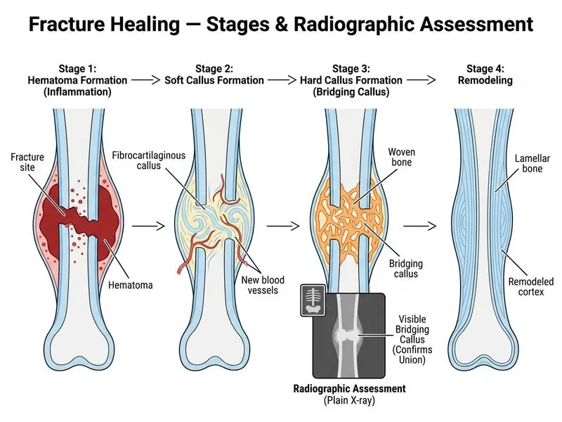
Plain radiography remains the investigation of choice for assessing fracture healing and identifying the stage of callus formation (soft callus, hard callus, and remodeling phases).
High-YieldNEET PG
At 6 weeks post-fracture, bridging callus formation is expected and is best visualized on plain radiographs. The presence of bridging callus (both endosteal and periosteal) indicates progression from the soft callus stage (weeks 2–3) to the hard callus stage (weeks 3–8).
Stages of Fracture Healing and Radiographic Findings
Table
Stage
Timeline
Radiographic Finding
Callus Type
Inflammatory
0–3 days
Fracture line, soft tissue swelling
None
Soft Callus
2–3 weeks
Fuzzy callus around fracture site
Periosteal + endosteal
Hard Callus
3–8 weeks
Bridging callus (continuous bone bridge)
Woven bone
Remodeling
8 weeks–1 year
Progressive mineralization, restoration of medullary canal
Lamellar bone
Clinical Pearl
Bridging callus on plain radiography is the clinical and radiological sign of fracture union. It indicates that the fracture has progressed beyond the soft callus stage and can now bear load.
Mnemonic: SHBR — Soft callus → Hard callus → Bridging → Remodeling (stages in order of appearance).
Why Other Investigations Are Inferior
CT scan: Unnecessary at this stage; reserved for complex fractures, intra-articular involvement, or when plain films are inconclusive. Adds radiation without additional diagnostic benefit for simple fracture healing assessment.
MRI: Not the investigation of choice for fracture healing; poor visualization of cortical bone and callus mineralization. Better suited for soft tissue injury assessment.
Ultrasound: Operator-dependent; less reliable for assessing bony callus maturity and bridging compared to radiography.
Rockwood & Green's Fractures in Adults, Ch 1
Loading illustration…
Practice similar questions
Sign up free to access AI-powered MCQ practice with detailed explanations and adaptive learning.