| Feature | Inflammatory Phase | Reparative (Callus) Phase | Remodeling Phase |
|---|---|---|---|
| Duration | 0–3 weeks | 3–12 weeks | Months to years |
| Dominant cells | Neutrophils, macrophages, RBCs | Fibroblasts, osteoblasts, chondrocytes | Osteocytes, osteoclasts |
| Tissue type | Blood clot, granulation tissue | Woven bone, cartilage (soft callus → hard callus) | Lamellar bone, cortical remodeling |
| Vascularity | Hemorrhage, edema | Neovascularization | Normalized |
| Radiographic appearance | Fracture line visible, no callus | Callus bridging fracture site | Callus resorption, cortical restoration |
The inflammatory phase is characterized by a cellular infiltrate dominated by polymorphonuclear leukocytes (neutrophils) and macrophages, with minimal osteogenic activity. This phase is essential for debridement of dead tissue and release of growth factors, but does NOT produce new bone.
The transition from inflammatory to reparative phase occurs around 3 weeks post-fracture. At this point, the fracture site shifts from a hemorrhagic/inflammatory milieu to an osteogenic environment where fibroblasts and osteoblasts proliferate and begin laying down woven bone (soft callus formation).
Neutrophils and macrophages = inflammatory phase; fibroblasts and osteoblasts = reparative phase. This is the single most reliable discriminator on exams.
INFLAMATORY = INFlammatory cells (Neutrophils, macrophages); REPAIR = REPair cells (fibroblasts, osteoblasts).
Rockwood & Green's Fractures in Adults Ch 1
Loading illustration…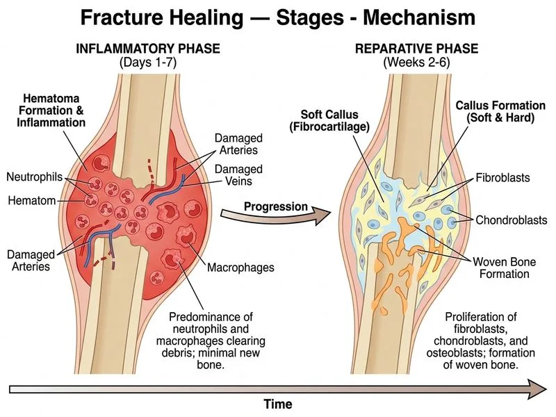
Sign up free to access AI-powered MCQ practice with detailed explanations and adaptive learning.
Daily MCQs, study tips, and topper strategies on Telegram.
Join on Telegram →