NEETPGAI
FeaturesNEET PGFMGEINI-CETBlogPricing
Log inStart Free
NEETPGAI

AI-powered NEET PG preparation platform. Master all 19 subjects with adaptive MCQs, AI tutoring, and spaced repetition.

Product

  • Features
  • Subjects
  • Previous Year Questions
  • NEET PG Preparation
  • FMGE Preparation
  • INI-CET Preparation
  • Compare
  • Pricing
  • Blog

Features

  • Adaptive MCQ Practice
  • AI Tutor
  • Mock Tests
  • Spaced Repetition

Resources

  • Blog
  • Study Guides
  • NEET PG Updates
  • Contact & support

Legal

  • Privacy Policy
  • Terms of Service

Stay updated

© 2026 NEETPGAI. All rights reserved.
    Subjects/Surgery/Head Injury — GCS and Management
    Head Injury — GCS and Management
    medium
    scissors Surgery

    A 35-year-old woman is admitted with a severe head injury (GCS 8/15) following a fall from height. Initial non-contrast CT head shows a right acute subdural hematoma with 8 mm midline shift. She is intubated and sedated. Her pupils are equal and reactive. Six hours later, her left pupil becomes dilated and unreactive. What is the most appropriate next investigation to assess the progression of injury?

    A. Electroencephalography (EEG)
    B. Intracranial pressure monitoring
    C. Transcranial Doppler ultrasound
    D. Repeat non-contrast CT head

    Explanation

    Investigation of Choice in Deteriorating Head Injury

    Key Point
    Repeat non-contrast CT head is the most appropriate investigation when there is clinical deterioration (anisocoria, change in GCS, pupillary changes) in a patient with known intracranial injury. It provides rapid assessment of hematoma expansion, herniation, and need for emergency neurosurgical intervention.
    Clinical Context: Herniation Syndrome

    Anisocoria (dilated left pupil) in a patient with right subdural hematoma suggests uncal herniation with compression of the ipsilateral oculomotor nerve (CN III). This is a neurosurgical emergency requiring immediate imaging to confirm hematoma expansion and guide surgical evacuation.

    Loading diagram...
    Why Repeat CT is Superior
    Table
    InvestigationRole in DeteriorationSpeedSpecificity
    Repeat CT headConfirms hematoma expansion, herniation, need for surgeryImmediateHighest
    TCD ultrasoundAssesses cerebral blood flow, not diagnostic for hematoma10–15 minLow for acute bleed
    ICP monitoringMeasures pressure, does not identify causeContinuousIndirect
    EEGDetects seizures, not diagnostic for hematoma30+ minNot relevant
    High-YieldNEET PG
    In a patient with known intracranial hemorrhage and acute neurological deterioration (especially pupillary changes), repeat CT is mandatory before any other investigation. It directly answers the question: "Is the hematoma expanding and causing herniation?"
    Clinical Pearl

    Anisocoria (unequal pupils) is a red flag for uncal herniation. The dilated pupil is on the side of the mass (ipsilateral). This patient requires emergent CT and neurosurgical consultation—not just monitoring.

    Warning
    Do not waste time with TCD, EEG, or ICP monitoring when there is clinical evidence of herniation. Repeat imaging is the priority.

    Loading illustration…Head Injury — GCS and Management diagram

    Practice similar questions

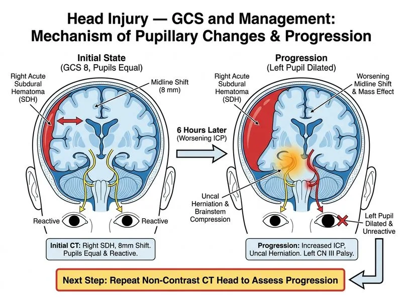
    Sign up free to access AI-powered MCQ practice with detailed explanations and adaptive learning.

    Start Practicing Free More Surgery Questions

    Join our NEET PG community

    Daily MCQs, study tips, and topper strategies on Telegram.

    Join on Telegram →