## Imaging Diagnosis of HCC in Cirrhosis ### Hallmark Enhancement Pattern **Key Point:** The pathognomonic imaging feature of HCC in a cirrhotic liver is **arterial phase hyperenhancement (APHE) followed by washout** in the portal venous or delayed phase. This pattern reflects the dual blood supply shift in HCC — increased hepatic artery contribution and loss of portal venous inflow. ### Mechanism Behind the Pattern In cirrhotic livers: - Normal hepatocytes receive 75% blood from portal vein, 25% from hepatic artery - Early HCC nodules develop increased arterial supply (angiogenesis) while retaining some portal venous flow - As HCC progresses, portal venous supply diminishes → arterial dominance - The washout occurs because HCC lacks the reticuloendothelial system present in normal liver ### Diagnostic Criteria (AASLD/EASL) | Nodule Size | Diagnostic Criteria | |---|---| | >20 mm | APHE + washout on one imaging modality (CT/MRI) | | 10–20 mm | APHE + washout on both CT AND MRI, OR biopsy | | <10 mm | Follow-up imaging (too small for reliable diagnosis) | **High-Yield:** Nodules showing APHE alone (without washout) are **not diagnostic** of HCC — they require either a second imaging modality or biopsy confirmation. ### Why Other Patterns Are Non-Specific - **Homogeneous arterial enhancement** alone occurs in both HCC and benign hyperenhancing nodules (adenomatous hyperplasia) - **Iso-attenuation** is typical of regenerative nodules and is reassuring - **Rim enhancement** suggests advanced HCC or cholangiocarcinoma, not early HCC **Clinical Pearl:** In a cirrhotic patient with a 15 mm nodule showing APHE on CT, you MUST obtain MRI with hepatobiliary contrast (gadoxetate) to confirm washout before diagnosing HCC. [cite:Robbins 10e Ch 20] 
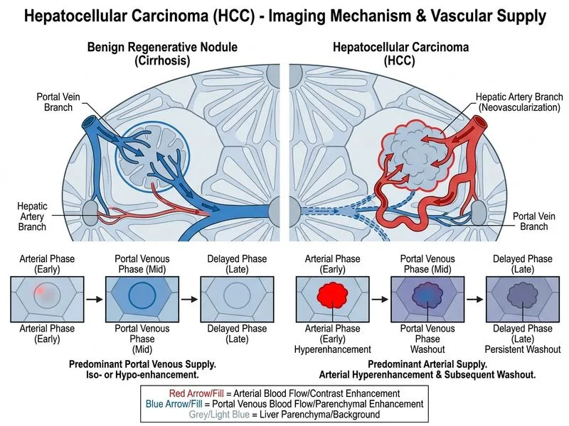
Sign up free to access AI-powered MCQ practice with detailed explanations and adaptive learning.