| Feature | Non-Bullous Impetigo | Bullous Impetigo |
|---|---|---|
| Lesion morphology | Honey-colored crusts on erythematous base | Flaccid bullae → erosions |
| Preceding vesicles | Absent; crusts form de novo | Present; blisters precede crusts |
| Causative organism | S. aureus (most common) | S. aureus (exfoliative toxin-producing strains) |
| Exotoxin involvement | No exotoxin; direct bacterial invasion | Exfoliative toxins (ETA, ETB) cause acantholysis |
| Frequency | 70% of impetigo cases | 30% of impetigo cases |
| Crusts appearance | Thick, adherent, honey/golden color | Thin, flaccid; erosions rather than crusts |
The hallmark of non-bullous impetigo is the formation of honey-colored crusts WITHOUT preceding fluid-filled vesicles. This occurs due to direct bacterial invasion of the epidermis, not exotoxin-mediated acantholysis.
Bullous impetigo is caused by exfoliative toxin-producing S. aureus strains (typically phage group II). These toxins cleave desmoglein-1 in the granular layer, creating intraepidermal acantholysis and flaccid bullae. Non-bullous impetigo lacks this exotoxin mechanism.
Non-bullous impetigo accounts for ~70% of all impetigo cases in clinical practice. The honey-crust without preceding vesicle is the single best discriminating feature and is pathognomonic for this variant.
HONEY = Non-bullous (Honey-colored crusts, direct invasion, NO exotoxin) BULLA = Bullous (Blisters precede crusts, exotoxin-mediated acantholysis)
Loading illustration…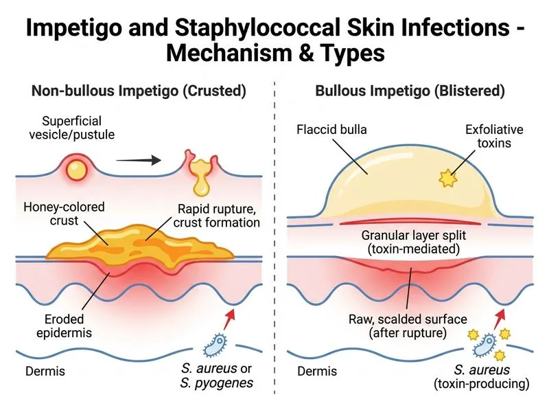
Sign up free to access AI-powered MCQ practice with detailed explanations and adaptive learning.
Daily MCQs, study tips, and topper strategies on Telegram.
Join on Telegram →