NEETPGAI
FeaturesNEET PGFMGEINI-CETBlogPricing
Log inStart Free
NEETPGAI

AI-powered NEET PG preparation platform. Master all 19 subjects with adaptive MCQs, AI tutoring, and spaced repetition.

Product

  • Features
  • Subjects
  • Previous Year Questions
  • NEET PG Preparation
  • FMGE Preparation
  • INI-CET Preparation
  • Compare
  • Pricing
  • Blog

Features

  • Adaptive MCQ Practice
  • AI Tutor
  • Mock Tests
  • Spaced Repetition

Resources

  • Blog
  • Study Guides
  • NEET PG Updates
  • Contact & support

Legal

  • Privacy Policy
  • Terms of Service

Stay updated

© 2026 NEETPGAI. All rights reserved.
    Subjects/Pathology/Lung Cancer — Non-Small Cell
    Lung Cancer — Non-Small Cell
    medium
    microscope Pathology

    Squamous cell carcinoma of the lung most commonly arises from which anatomical location within the bronchial tree?

    A. Alveolar ducts and alveoli
    B. Segmental and subsegmental bronchi
    C. Lobar and main bronchi
    D. Terminal bronchioles

    Explanation

    Anatomical Origin of Lung Squamous Cell Carcinoma

    Key Point
    Squamous cell carcinoma (SCC) of the lung arises from the proximal airways — lobar and main bronchi — where the respiratory epithelium undergoes squamous metaplasia in response to chronic irritation (smoking).
    Origin & Pathogenesis
    1. 1.
      Chronic smoking → irritation of proximal bronchial epithelium
    2. 2.
      Squamous metaplasia → replacement of normal ciliated columnar epithelium with stratified squamous epithelium
    3. 3.
      Dysplasia → low-grade → high-grade dysplasia
    4. 4.
      Carcinoma → invasive squamous cell carcinoma
    Anatomical Predilection
    • Lobar and main bronchi (central airways) — most common site
    • Hilar location → central lung mass on imaging
    • Proximal location makes it accessible to bronchoscopy for diagnosis
    • Contrast: adenocarcinoma arises in peripheral lung (distal airways, alveoli)
    Clinical Implications
    Table
    FeatureSquamous Cell CarcinomaAdenocarcinoma
    LocationCentral (lobar/main bronchi)Peripheral (distal airways)
    SmokingStrongly associatedWeakly associated
    MetaplasiaSquamousMucous/glandular
    CavitationCommonRare
    BronchoscopyAccessibleOften not visible
    High-YieldNEET PG
    SCC = central hilar mass in a smoker. This is a classic imaging-pathology correlation tested in NEET PG.
    Clinical Pearl
    Central location and accessibility to bronchoscopy make SCC easier to diagnose histologically compared to peripheral adenocarcinoma, which often requires CT-guided biopsy or resection specimen.

    Loading illustration…Lung Cancer — Non-Small Cell diagram

    Practice similar questions

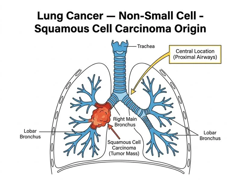
    Sign up free to access AI-powered MCQ practice with detailed explanations and adaptive learning.

    Start Practicing Free More Pathology Questions

    Join our NEET PG community

    Daily MCQs, study tips, and topper strategies on Telegram.

    Join on Telegram →