A 58-year-old male smoker (40 pack-years) presents with persistent cough and haemoptysis for 3 weeks. Chest X-ray shows a 4 cm peripheral nodule in the right lower lobe with irregular borders. CT chest reveals no mediastinal lymphadenopathy. Bronchoscopy with biopsy shows malignant cells arranged in nests with prominent desmoplastic stromal reaction and keratinization. What is the most likely histological type of lung cancer?
A. Large cell carcinoma
B. Squamous cell carcinoma
C. Small cell carcinoma
D. Adenocarcinoma
Explanation
Histopathological Diagnosis
Key Point
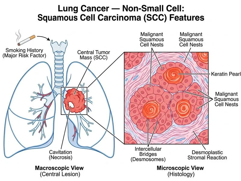
The combination of keratinization and desmoplastic stromal reaction (fibrous tissue response) is pathognomonic for squamous cell carcinoma of the lung.
Clinical & Pathological Features of Squamous Cell Carcinoma
Table
Feature
Squamous Cell
Adenocarcinoma
Large Cell
Small Cell
Keratinization
Present (hallmark)
Absent
Absent
Absent
Desmoplasia
Prominent
Mild
Absent
Absent
Smoking association
Very strong (90%)
Moderate
Strong
Very strong
Location
Central/hilar
Peripheral
Peripheral
Central
Bronchoscopy finding
Endobronchial lesion
Distal/parenchymal
Rare
Central
Pathological Architecture
High-YieldNEET PG
Squamous cell carcinoma shows:
1.
Nests of polygonal cells with clear cytoplasm
2.
Intercellular bridges (desmosomes) — sign of squamous differentiation
3.
Keratin pearl formation (concentric layers of keratinized cells)
4.
Desmoplastic response (abundant collagen deposition by fibroblasts)
Clinical Context
Clinical Pearl
This patient's presentation is classic for squamous cell carcinoma:
Heavy smoking history (40 pack-years)
Central/hilar location tendency (though can be peripheral)
Haemoptysis (from endobronchial ulceration)
Desmoplastic stromal reaction visible on histology
Do not confuse squamous cell carcinoma with adenocarcinoma. Adenocarcinoma shows mucin production and glandular differentiation, NOT keratinization. Large cell carcinoma lacks both keratinization and glandular features — it is a diagnosis of exclusion.
Loading illustration…
Practice similar questions
Sign up free to access AI-powered MCQ practice with detailed explanations and adaptive learning.