NEETPGAI
FeaturesNEET PGFMGEINI-CETBlogPricing
Log inStart Free
NEETPGAI

AI-powered NEET PG preparation platform. Master all 19 subjects with adaptive MCQs, AI tutoring, and spaced repetition.

Product

  • Features
  • Subjects
  • Previous Year Questions
  • NEET PG Preparation
  • FMGE Preparation
  • INI-CET Preparation
  • Compare
  • Pricing
  • Blog

Features

  • Adaptive MCQ Practice
  • AI Tutor
  • Mock Tests
  • Spaced Repetition

Resources

  • Blog
  • Study Guides
  • NEET PG Updates
  • Contact & support

Legal

  • Privacy Policy
  • Terms of Service

Stay updated

© 2026 NEETPGAI. All rights reserved.
    Subjects/Pathology/Lymphomas — Non-Hodgkin
    Lymphomas — Non-Hodgkin
    medium
    microscope Pathology

    A 58-year-old man from Delhi presents with a 3-month history of progressive lymphadenopathy, fever, and night sweats. Physical examination reveals generalized lymphadenopathy and hepatosplenomegaly. Laboratory investigations show hemoglobin 9.2 g/dL, WBC 4800/μL with 15% atypical lymphocytes, and LDH 680 U/L. A chest X-ray shows mediastinal widening. What is the most appropriate next step in management?

    A. Initiate empirical broad-spectrum antibiotics and repeat investigations in 2 weeks
    B. Start rituximab-based chemotherapy after clinical staging
    C. Excisional lymph node biopsy with flow cytometry and immunohistochemistry
    D. Perform CT chest and abdomen followed by bone marrow biopsy

    Explanation

    Clinical Context

    This patient presents with B symptoms (fever, night sweats), lymphadenopathy, hepatosplenomegaly, elevated LDH, and mediastinal involvement — a classic presentation suggestive of non-Hodgkin lymphoma (NHL), likely a high-grade subtype given the constitutional symptoms.

    Diagnostic Hierarchy in NHL

    Key Point
    Tissue diagnosis is mandatory before any treatment in NHL. The diagnosis cannot be established on clinical features, imaging, or laboratory findings alone.
    High-YieldNEET PG
    Excisional (or core needle) biopsy with immunophenotyping (flow cytometry) and immunohistochemistry is the gold standard first step. This provides:
    • Histologic subtype (diffuse large B-cell, Burkitt, lymphoblastic, etc.)
    • Immunophenotype (B-cell vs. T-cell, CD5+/− status)
    • Prognostic markers (Ki-67, TP53 status in some cases)

    Why Excisional Biopsy?

    Fine-needle aspiration (FNA) is inadequate for lymphoma diagnosis because it cannot assess tissue architecture, which is essential for subtype classification. Excisional biopsy of the most accessible lymph node (avoiding small nodes <1 cm) is preferred.

    Staging Comes After Diagnosis

    Once histology confirms NHL, staging investigations (CT, PET-CT, bone marrow biopsy if indicated) follow to determine extent of disease and prognosis — but only after tissue diagnosis is secured.

    Clinical Pearl
    Mediastinal involvement in a young patient with B symptoms and elevated LDH suggests primary mediastinal B-cell lymphoma (PMBL) or Burkitt lymphoma — both requiring urgent tissue diagnosis and risk stratification before treatment initiation.

    Loading illustration…Lymphomas — Non-Hodgkin diagram

    Practice similar questions

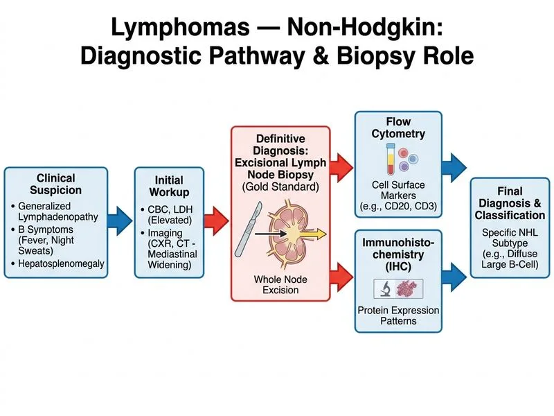
    Sign up free to access AI-powered MCQ practice with detailed explanations and adaptive learning.

    Start Practicing Free More Pathology Questions

    Join our NEET PG community

    Daily MCQs, study tips, and topper strategies on Telegram.

    Join on Telegram →