Granulation tissue formation; macrophage infiltration and phagocytosis
3–8 weeks
Fibrosis and scar formation
High-YieldNEET PG
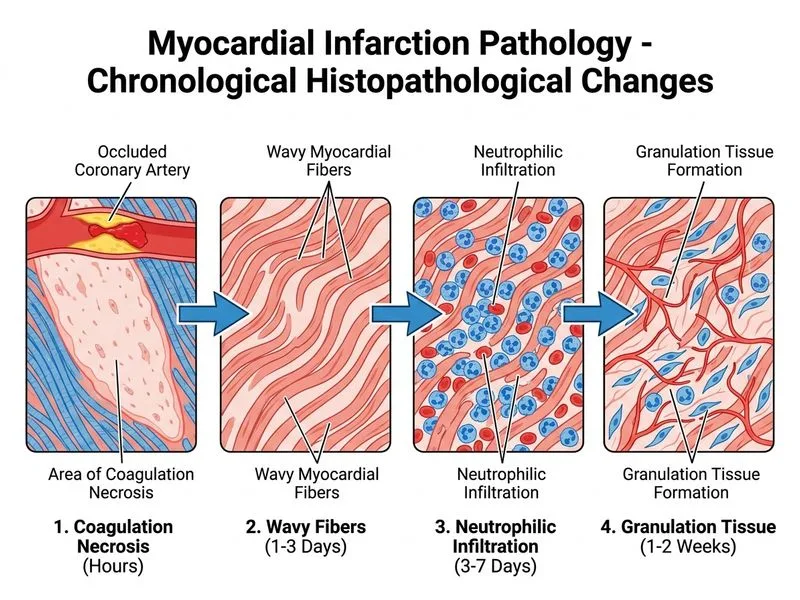
The earliest light microscopic change in acute MI is wavy fibers — non-contractile dead myocytes at the border zone are passively stretched and buckled by the pull of adjacent viable myocytes, producing a wavy appearance visible within 1–4 hours. Coagulation necrosis (hypereosinophilia, nuclear loss) becomes established by 4–12 hours. Neutrophilic infiltration follows after 12–24 hours as the acute inflammatory response is mounted. Granulation tissue appears after approximately 1 week as healing begins.
Per Robbins Basic Pathology (10th ed.), wavy fibers are the hallmark of the earliest (1–4 hour) phase of MI visible on H&E staining. The presence of neutrophils is a useful marker to date an infarction to >12 hours old. Absence of neutrophils in a necrotic area suggests infarction <12 hours old.