NEETPGAI
FeaturesNEET PGFMGEINI-CETBlogPricing
Log inStart Free
NEETPGAI

AI-powered NEET PG preparation platform. Master all 19 subjects with adaptive MCQs, AI tutoring, and spaced repetition.

Product

  • Features
  • Subjects
  • Previous Year Questions
  • NEET PG Preparation
  • FMGE Preparation
  • INI-CET Preparation
  • Compare
  • Pricing
  • Blog

Features

  • Adaptive MCQ Practice
  • AI Tutor
  • Mock Tests
  • Spaced Repetition

Resources

  • Blog
  • Study Guides
  • NEET PG Updates
  • Contact & support

Legal

  • Privacy Policy
  • Terms of Service

Stay updated

© 2026 NEETPGAI. All rights reserved.
    Subjects/Pathology/Myocardial Infarction Pathology
    Myocardial Infarction Pathology
    medium
    microscope Pathology

    A 58-year-old man with hypertension and diabetes presents with acute chest pain and ST elevation in leads II, III, and aVF. Coronary angiography shows complete occlusion of the right coronary artery. Which pathological feature best distinguishes transmural infarction from subendocardial infarction in this case?

    A. Involvement of the full thickness of the ventricular wall including the epicardium
    B. Absence of neutrophilic infiltration at the border zone
    C. Presence of coagulation necrosis extending from endocardium to epicardium
    D. Isolated necrosis limited to the inner third of the myocardium

    Explanation

    Transmural vs. Subendocardial Infarction: Pathological Distinction

    The depth and extent of myocardial necrosis fundamentally distinguish these two patterns and correlate with the clinical presentation and prognosis.

    Comparative Pathology
    Table
    FeatureTransmural MISubendocardial MI
    Depth of necrosisFull thickness (endocardium to epicardium)Inner third of myocardium only
    Typical causeComplete coronary occlusionSevere stenosis, demand ischemia, or reperfusion
    ECG findingST elevation + pathological Q wavesST depression, T-wave inversion (no Q waves)
    Complication riskHigh (rupture, aneurysm, VSD)Lower
    Epicardial involvementYes (defines transmural)No
    Pericarditis riskHigh (transmural necrosis irritates pericardium)Low
    Key Point:

    Full-thickness involvement including the epicardium is the defining pathological criterion for transmural MI. In this case, complete RCA occlusion (inferior STEMI) typically causes transmural necrosis in the inferior wall.

    Clinical Pearl:

    Transmural MI carries higher risk of mechanical complications (free wall rupture, ventricular septal defect, papillary muscle rupture) because the necrosis extends through all layers, weakening the structural integrity of the wall.

    High-Yield:

    Remember the depth rule: Transmural = full thickness (endocardium + epicardium); Subendocardial = inner third only. This distinction is testable in both pathology and clinical reasoning questions.

    Mnemonic:

    TRANSMURAL = THROUGH-ALL — necrosis extends through all layers of the myocardium.

    Loading illustration…Myocardial Infarction Pathology diagram

    Practice similar questions

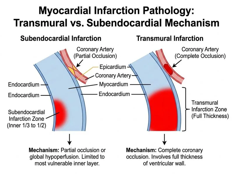
    Sign up free to access AI-powered MCQ practice with detailed explanations and adaptive learning.

    Start Practicing Free More Pathology Questions

    Join our NEET PG community

    Daily MCQs, study tips, and topper strategies on Telegram.

    Join on Telegram →