
## Clinical Presentation Analysis **Key Point:** Painless jaundice with Courvoisier sign (palpable, non-tender gallbladder) strongly suggests malignant obstruction of the CBD — most commonly pancreatic head carcinoma. **Clinical Pearl:** The combination of dilated intrahepatic and extrahepatic bile ducts with a dilated pancreatic duct ("double duct sign") on ultrasound is highly suggestive of pancreatic head malignancy. No stone is visible, making choledocholithiasis unlikely. ## Why CT Abdomen with IV Contrast Is the Answer In a patient with suspected pancreatic head malignancy presenting with obstructive jaundice, **contrast-enhanced CT (CECT) of the abdomen and pelvis** is the most appropriate next imaging modality after ultrasound. This is supported by major guidelines (NCCN, ACR, and standard surgical oncology practice): 1. **Characterizes the mass**: CECT identifies the pancreatic head lesion, its size, and morphology with high sensitivity. 2. **Assesses resectability**: Evaluates vascular involvement (SMA, SMV, portal vein, celiac axis) — the critical determinant for surgical planning. 3. **Detects metastases**: Identifies liver metastases, peritoneal deposits, and lymphadenopathy. 4. **Guides further management**: Determines whether the patient proceeds to surgery, neoadjuvant therapy, or palliative intervention. | Imaging Modality | Role in This Scenario | Advantage | Limitation | |---|---|---|---| | **CT abdomen/pelvis (IV contrast)** | **First-line after US for suspected pancreatic malignancy** | Mass characterization, vascular involvement, staging, resectability | Less sensitive for very small lesions | | MRCP | Ductal anatomy, biliary stricture characterization | Non-invasive, excellent ductal detail | Does not assess resectability or vascular involvement as well as CT | | EUS | Tissue diagnosis (FNA), small lesion detection | Highest sensitivity for small lesions | Operator-dependent, invasive, not for staging | | HIDA scan | Biliary dyskinesia, cystic duct patency | Functional assessment | Poor spatial resolution, not indicated for obstruction workup | ## Imaging Algorithm for Suspected Pancreatic Head Malignancy - **Step 1**: Ultrasound (confirms ductal dilation, rules out stones) ✓ Done - **Step 2**: **CECT abdomen/pelvis** — establishes diagnosis, assesses resectability - **Step 3**: EUS ± FNA if tissue diagnosis needed before surgery or if CT is inconclusive - **Step 4**: MRCP if ductal anatomy needs further delineation (e.g., pre-ERCP planning) **High-Yield (Harrison's Principles of Internal Medicine / Sabiston Textbook of Surgery):** For suspected periampullary or pancreatic head malignancy, CECT is the investigation of choice after ultrasound. It provides the most comprehensive information for diagnosis AND surgical planning in a single study. MRCP is complementary for ductal anatomy but does not replace CT for staging and resectability assessment. **Key Point:** Courvoisier sign (palpable non-tender gallbladder in jaundice) indicates malignant obstruction of CBD. The next step is CECT abdomen — not MRCP — because the clinical priority is to characterize the mass and determine resectability, not merely to delineate ductal anatomy. 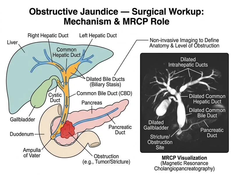
Sign up free to access AI-powered MCQ practice with detailed explanations and adaptive learning.