NEETPGAI
FeaturesNEET PGFMGEINI-CETBlogPricing
Log inStart Free
NEETPGAI

AI-powered NEET PG preparation platform. Master all 19 subjects with adaptive MCQs, AI tutoring, and spaced repetition.

Product

  • Features
  • Subjects
  • Previous Year Questions
  • NEET PG Preparation
  • FMGE Preparation
  • INI-CET Preparation
  • Compare
  • Pricing
  • Blog

Features

  • Adaptive MCQ Practice
  • AI Tutor
  • Mock Tests
  • Spaced Repetition

Resources

  • Blog
  • Study Guides
  • NEET PG Updates
  • Contact & support

Legal

  • Privacy Policy
  • Terms of Service

Stay updated

© 2026 NEETPGAI. All rights reserved.
    Subjects/Ophthalmology/Orbital Cellulitis
    Orbital Cellulitis
    hard
    eye Ophthalmology

    A 28-year-old woman from Bangalore with acute orbital cellulitis secondary to ethmoid sinusitis has been on IV ceftriaxone and vancomycin for 5 days. She shows clinical improvement (reduced proptosis, improved eye movements), and repeat imaging shows no abscess. However, her fever persists and she complains of severe headache. Blood cultures from admission grew Streptococcus pneumoniae sensitive to cephalosporins. What is the most appropriate next step?

    A. Continue IV antibiotics; repeat imaging to exclude abscess formation or cavernous sinus thrombosis
    B. Discontinue vancomycin and continue ceftriaxone monotherapy; observe for 48 hours
    C. Continue current IV antibiotics for a total of 2–4 weeks; add lumbar puncture to exclude meningitis given persistent fever and headache
    D. Switch to oral cephalosporin immediately to complete outpatient therapy

    Explanation

    Clinical Scenario Analysis

    This patient has orbital cellulitis with clinical improvement on appropriate IV antibiotics, but persistent fever and new/worsening headache raise concern for a serious complication: cavernous sinus thrombosis (CVT) or occult abscess. Despite initial imaging showing no abscess, complications can develop during treatment.

    Differential Diagnosis of Persistent Fever in Orbital Cellulitis

    Table
    ComplicationClinical FeaturesImaging FindingsManagement
    Cavernous sinus thrombosisBilateral signs, headache, altered mental status, seizuresContrast-enhanced MRI: filling defect in cavernous sinusContinue/escalate IV antibiotics, consider anticoagulation
    Orbital abscessFailure to improve, localized pain, proptosis plateauCT/MRI: loculated collectionIV antibiotics + surgical drainage
    MeningitisHeadache, neck stiffness, photophobia, altered sensoriumCSF pleocytosis, elevated proteinIV antibiotics + lumbar puncture after imaging
    Inadequate source controlPersistent sinusitis signsImaging: active sinusitis, fluid levelsENT consultation, possible sinus drainage

    Key Management Principle

    High-YieldNEET PG
    Persistent fever and new headache in a patient with orbital cellulitis on appropriate antibiotics is a red flag for complications (especially cavernous sinus thrombosis). Repeat imaging is mandatory to exclude abscess and assess for CVT before considering de-escalation or discharge.
    Key Point
    Cavernous sinus thrombosis is a life-threatening complication with mortality 5–10% even with treatment. Early detection via imaging and continued aggressive IV antibiotics are critical. Lumbar puncture is performed after imaging excludes mass effect.
    Clinical Pearl
    Headache in orbital cellulitis may reflect:
    1. 1.
      Sinus inflammation (expected)
    2. 2.
      Early meningitis (rare, but serious)
    3. 3.
      Cavernous sinus thrombosis (most concerning in this context)

    Imaging (contrast-enhanced MRI with venography) is more sensitive than lumbar puncture for detecting CVT.

    Why Repeat Imaging?

    • Initial imaging was done at presentation; complications can develop during treatment
    • MRI with contrast and venography is optimal for detecting cavernous sinus involvement
    • Guides decision on duration of IV therapy, need for anticoagulation, and surgical intervention

    Antibiotic Continuation

    The organism (S. pneumoniae) is sensitive to cephalosporins, so ceftriaxone monotherapy is reasonable after complications are excluded and clinical improvement is documented. However, vancomycin should be continued until imaging excludes CVT and lumbar puncture (if performed) excludes meningitis.

    Timeline for De-escalation

    • Continue IV antibiotics for 2–4 weeks total
    • Switch to oral antibiotics only after:
      • Imaging excludes complications
      • Clinical improvement is sustained (48–72 hours afebrile)
      • Organism susceptibilities confirm oral agent efficacy

    Loading illustration…Orbital Cellulitis diagram

    Practice similar questions

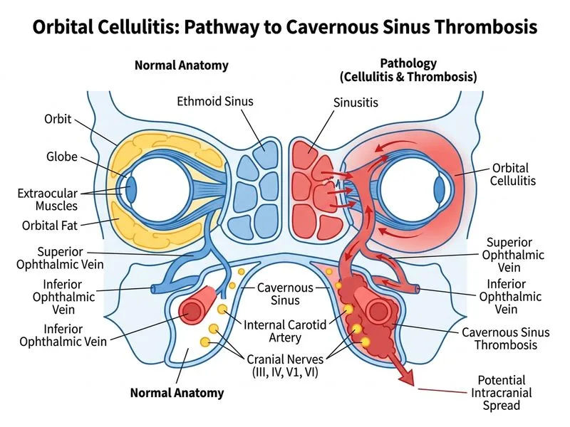
    Sign up free to access AI-powered MCQ practice with detailed explanations and adaptive learning.

    Start Practicing Free More Ophthalmology Questions

    Join our NEET PG community

    Daily MCQs, study tips, and topper strategies on Telegram.

    Join on Telegram →