## Clinical Diagnosis: Otosclerosis ### Key Clinical Features **Key Point:** Otosclerosis is a primary bone dysplasia of the otic capsule causing progressive fixation of the stapes footplate, leading to conductive hearing loss. ### Diagnostic Criteria Met in This Case | Feature | Finding | Significance | |---------|---------|---------------| | Age of onset | 32 years (young adult) | Typical age 20–40 years; rare before age 15 | | Gender | Female | 2:1 female predominance | | Progression | Bilateral, gradual | Bilateral involvement in ~70% of cases | | Hearing pattern | Conductive (air-bone gap 20 dB) | Stapes fixation reduces ossicular mobility | | Weber test | Bone conduction > air conduction | Confirms conductive loss | | Otoscopy | Normal TM | Rules out middle ear infection | | CT findings | Bilateral stapes fixation, normal ossicles | Pathognomonic for otosclerosis | | Associated symptoms | Tinnitus, vertigo | Common in otosclerosis | ### Pathophysiology 1. **Abnormal bone remodeling** in the otic capsule (endochondral bone) 2. **Stapes footplate fixation** at the oval window 3. **Reduced ossicular chain mobility** → conductive hearing loss 4. Progression may lead to **mixed or sensorineural hearing loss** (cochlear involvement) ### High-Yield Mnemonic: **OTOSCLEROSIS** - **O**tic capsule dysplasia - **T**ypical age 20–40 years - **O**ssicular fixation (stapes) - **S**ensorineural + conductive loss possible - **C**arhart notch (bone conduction peak at 2 kHz on audiometry) - **L**ateral canal (anterior oval window involvement) - **E**ndochondral bone remodeling - **R**adiolucent foci on HRCT (lytic phase) - **O**ssicles normal on imaging - **S**urgical cure: stapedectomy/stapedotomy - **I**nheritance: autosomal dominant (60% of cases) - **S**ymmetrical bilateral involvement common ### Carhart Sign **Clinical Pearl:** A characteristic **bone conduction peak at 2 kHz** (Carhart notch) on audiometry is highly suggestive of otosclerosis, though not pathognomonic. This represents the resonance frequency of the ossicular chain when stapes is fixed. ### Imaging Findings ```mermaid flowchart TD A[Otosclerosis on HRCT]:::outcome --> B{Phase of disease}:::decision B -->|Lytic phase| C[Radiolucent foci around oval window]:::action B -->|Mixed phase| D[Both radiolucent and sclerotic areas]:::action B -->|Sclerotic phase| E[Sclerotic bone, stapes fixation]:::action C --> F[Stapes footplate fixation]:::outcome D --> F E --> F F --> G[Conductive hearing loss]:::outcome ``` ### Natural History **High-Yield:** Progressive hearing loss occurs in ~2/3 of patients; ~1/3 remain stable. Pregnancy may accelerate progression (hormonal factors). ### Management - **Hearing aids** for mild-to-moderate loss - **Stapedectomy or stapedotomy** (gold standard surgical treatment) - Success rate: >90% restoration of air-bone gap - Risks: sensorineural hearing loss, facial nerve injury, perilymphatic fistula - **Sodium fluoride** (experimental; evidence weak) — may slow progression in some cases [cite:Dhingra ENT 8e Ch 11] 
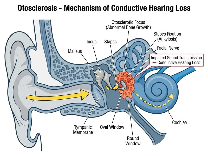
Sign up free to access AI-powered MCQ practice with detailed explanations and adaptive learning.