Which of the following is the most common site of peptic ulcer disease in the duodenum?
A. Third part of duodenum (D3)
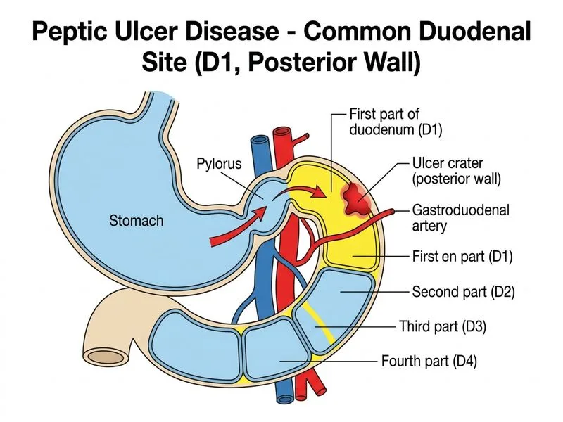
B. First part of duodenum (D1), posterior wall
C. Second part of duodenum (D2)
D. First part of duodenum (D1), anterior wall
Explanation
Anatomical Location of Duodenal Ulcers
Key Point
The anterior wall of the first part of the duodenum (D1) is the most common site of duodenal peptic ulcers. Approximately 95% of all duodenal ulcers occur in D1 (the duodenal bulb), and of these, the anterior wall is more frequently affected than the posterior wall.
Why the First Part, Anterior Wall?
1.
Direct acid exposure: The duodenal bulb (D1) receives the highest concentration of gastric acid immediately after it exits the pylorus, making it the most vulnerable segment.
2.
Anterior predominance: Standard surgical and pathology texts (Schwartz's Principles of Surgery, Robbins & Cotran) describe the anterior wall of D1 as the more common site for ulceration and perforation.
3.
Helicobacter pylori: The majority of duodenal ulcers are associated with H. pylori infection, which preferentially colonizes the gastric metaplastic epithelium of the duodenal bulb.
Clinical Significance of Anterior vs. Posterior Wall Ulcers
Table
Feature
Anterior Wall (D1)
Posterior Wall (D1)
Frequency
More common
Less common
Complication
Perforation → acute peritonitis
Erosion → hemorrhage (gastroduodenal artery)
Presentation
Sudden severe abdominal pain, board-like rigidity
Massive upper GI bleeding (hematemesis/melena)
Surgery
Omental patch (Graham patch)
Ligation of gastroduodenal artery
Clinical Pearl
Although posterior wall ulcers are classically associated with life-threatening hemorrhage (due to proximity of the gastroduodenal artery), the anterior wall is the more common site of ulceration overall. The mnemonic: "Anterior = Air under diaphragm (perforation); Posterior = Pulsatile bleeding (gastroduodenal artery)."
High-YieldNEET PG
For NEET PG/INI-CET, remember that ~95% of duodenal ulcers occur in D1 (duodenal bulb), with the anterior wall being the most common site. D2 ulcers are associated with Zollinger-Ellison syndrome (multiple/atypical ulcers beyond the bulb).
Reference: Robbins & Cotran Pathologic Basis of Disease, 10th ed., Chapter on Gastrointestinal Tract; Schwartz's Principles of Surgery, 11th ed.
Loading illustration…
Practice similar questions
Sign up free to access AI-powered MCQ practice with detailed explanations and adaptive learning.