A 35-year-old woman with known cystic fibrosis presents with acute dyspnea. Chest X-ray shows a right-sided pneumothorax. Which imaging finding on high-resolution CT best distinguishes a tension pneumothorax from a simple (non-tension) pneumothorax?
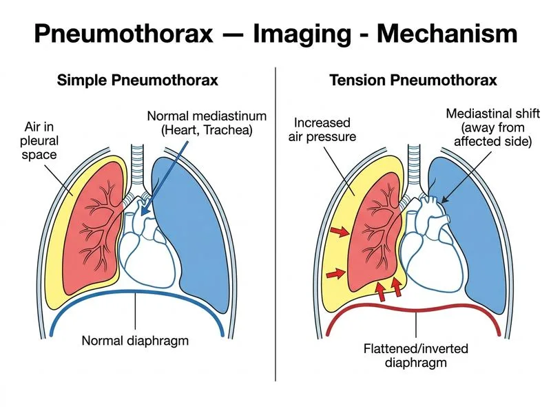
A. Mediastinal shift (deviation of mediastinum away from the side of pneumothorax) and flattening or inversion of the ipsilateral hemidiaphragm
B. Presence of a visible visceral pleural line separating collapsed lung from air-filled pleural space
C. Complete opacification of the hemithorax with absence of any visible lung parenchyma
D. Subcutaneous emphysema tracking along the chest wall and neck
Explanation
Tension vs. Non-Tension Pneumothorax: Imaging Discriminators
Critical Distinguishing Features
Key Point
Mediastinal shift (away from the pneumothorax side) and hemidiaphragmatic flattening or inversion are the hallmark imaging signs of tension pneumothorax and distinguish it from simple pneumothorax.
Pathophysiology Behind Imaging Findings
High-YieldNEET PG
In tension pneumothorax, positive intrapleural pressure accumulates during inspiration and fails to equilibrate during expiration. This causes:
1.
Progressive mediastinal displacement away from the affected side
2.
Compression of the contralateral lung
3.
Flattening or paradoxical inversion of the ipsilateral hemidiaphragm
In simple pneumothorax, intrapleural pressure remains subatmospheric or atmospheric; no progressive shift occurs.
Imaging Comparison Table
Table
Feature
Simple Pneumothorax
Tension Pneumothorax
Mediastinal position
Midline or minimal shift
Marked shift away from PTX side
Hemidiaphragm
Normal curvature
Flattened or inverted (pushed down)
Contralateral lung
Normal
Compressed
Pleural line
Visible, sharp
Visible but with mass effect
Clinical urgency
Managed conservatively or with aspiration
EMERGENCY — requires immediate decompression
Hemodynamic stability
Stable
Unstable (hypotension, shock)
Why Mediastinal Shift Is the Key Discriminator
Clinical Pearl
On frontal chest X-ray, measure the position of the mediastinum (trachea, heart) relative to the midline. In tension pneumothorax, the mediastinum is displaced away from the pneumothorax side by >2 cm. On CT, this shift is even more obvious and accompanied by hemidiaphragmatic changes.