NEETPGAI
FeaturesBlogComparePricing
Log inStart Free
NEETPGAI

AI-powered NEET PG preparation platform. Master all 19 subjects with adaptive MCQs, AI tutoring, and spaced repetition.

Product

  • Features
  • Subjects
  • Previous Year Questions
  • Compare
  • Pricing
  • Blog

Features

  • Adaptive MCQ Practice
  • AI Tutor
  • Mock Tests
  • Spaced Repetition

Resources

  • Blog
  • Study Guides
  • NEET PG Updates
  • Contact & support

Legal

  • Privacy Policy
  • Terms of Service

Stay updated

© 2026 NEETPGAI. All rights reserved.
    Subjects/Anatomy/Popliteal Fossa
    Popliteal Fossa
    medium
    bone Anatomy

    A 32-year-old male presents to the orthopaedic clinic with a popliteal cyst (Baker's cyst). During clinical examination, the physician palpates the popliteal fossa to assess for associated joint pathology. Which of the following structures forms the medial boundary of the popliteal fossa?

    A. Vastus medialis and adductor magnus
    B. Semitendinosus and semimembranosus
    C. Biceps femoris and common peroneal nerve
    D. Gracilis and sartorius

    Explanation

    ## Popliteal Fossa Boundaries The popliteal fossa is a diamond-shaped space located on the posterior aspect of the knee joint. Its boundaries are: **Medial boundary:** Semitendinosus and semimembranosus (superomedial) and medial head of gastrocnemius (inferomedial) **Lateral boundary:** Biceps femoris (superolateral) and lateral head of gastrocnemius (inferolateral) **Floor:** Popliteus muscle, posterior joint capsule, and oblique popliteal ligament **Roof:** Skin, fascia, and semitendinosus aponeurosis ## Key Point: **The medial boundary is formed by the semitendinosus and semimembranosus muscles superiorly**, which are the medial hamstring muscles. Inferiorly, the medial head of gastrocnemius completes the medial boundary. ## Clinical Pearl: Baker's cysts (popliteal cysts) arise from the joint capsule, typically between the medial head of gastrocnemius and semimembranosus. Understanding the anatomy is crucial for clinical examination and ultrasound assessment. ![Popliteal Fossa diagram](https://mmcphlazjonnzmdysowq.supabase.co/storage/v1/object/public/blog-images/explanation/2056.webp)

    Practice similar questions

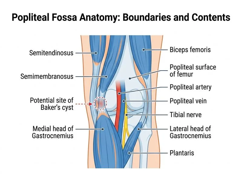
    Sign up free to access AI-powered MCQ practice with detailed explanations and adaptive learning.

    Start Practicing Free More Anatomy Questions