NEETPGAI
FeaturesBlogComparePricing
Log inStart Free
NEETPGAI

AI-powered NEET PG preparation platform. Master all 19 subjects with adaptive MCQs, AI tutoring, and spaced repetition.

Product

  • Features
  • Subjects
  • Previous Year Questions
  • Compare
  • Pricing
  • Blog

Features

  • Adaptive MCQ Practice
  • AI Tutor
  • Mock Tests
  • Spaced Repetition

Resources

  • Blog
  • Study Guides
  • NEET PG Updates
  • Contact & support

Legal

  • Privacy Policy
  • Terms of Service

Stay updated

© 2026 NEETPGAI. All rights reserved.
    Subjects/Anatomy/Popliteal Fossa
    Popliteal Fossa
    medium
    bone Anatomy

    A 45-year-old man undergoes surgical exploration of the popliteal fossa for removal of a thrombosed popliteal artery aneurysm. During dissection, the surgeon must carefully identify and preserve the tibial nerve. Which of the following statements correctly describes the anatomical relationship of the tibial nerve within the popliteal fossa?

    A. The tibial nerve lies deep to the popliteal vein and artery, and is the most medial of the three main neurovascular structures
    B. The tibial nerve lies superficial to the popliteal vein and crosses it medially at the level of the knee joint
    C. The tibial nerve runs along the lateral border of the popliteal fossa and is crossed by the common peroneal nerve
    D. The tibial nerve is the most superficial neurovascular structure in the popliteal fossa and lies posterior to the popliteal vein

    Explanation

    ## Anatomical Position of Tibial Nerve in Popliteal Fossa The popliteal fossa contains three main neurovascular structures arranged in a specific anatomical relationship: ### Layers (Superficial to Deep): 1. **Popliteal vein** — lies most superficially 2. **Popliteal artery** — lies deep to the vein 3. **Tibial nerve** — lies deepest, posterior to both vessels ### Medial-Lateral Arrangement: - **Medial**: Tibial nerve (most medial) - **Central**: Popliteal artery - **Lateral**: Common peroneal nerve (runs along lateral border) ### Clinical Significance: The tibial nerve's deep and medial position makes it vulnerable during surgical procedures in the popliteal fossa. During aneurysm repair or vascular surgery, careful dissection is required to avoid iatrogenic nerve injury, which would result in loss of ankle plantarflexion and toe flexion. **Key Point:** The mnemonic for popliteal fossa contents from superficial to deep is **VAT** (Vein, Artery, Tibial nerve), with the tibial nerve being the deepest and most medial structure. **High-Yield Fact:** The common peroneal nerve diverges from the sciatic nerve at the superior border of the popliteal fossa and runs along the lateral margin, making it the most lateral neurovascular element. ![Popliteal Fossa diagram](https://mmcphlazjonnzmdysowq.supabase.co/storage/v1/object/public/blog-images/explanation/2058.webp)

    Practice similar questions

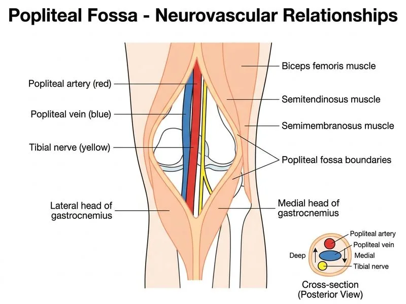
    Sign up free to access AI-powered MCQ practice with detailed explanations and adaptive learning.

    Start Practicing Free More Anatomy Questions