NEETPGAI
FeaturesNEET PGFMGEINI-CETBlogPricing
Log inStart Free
NEETPGAI

AI-powered NEET PG preparation platform. Master all 19 subjects with adaptive MCQs, AI tutoring, and spaced repetition.

Product

  • Features
  • Subjects
  • Previous Year Questions
  • NEET PG Preparation
  • FMGE Preparation
  • INI-CET Preparation
  • Compare
  • Pricing
  • Blog

Features

  • Adaptive MCQ Practice
  • AI Tutor
  • Mock Tests
  • Spaced Repetition

Resources

  • Blog
  • Study Guides
  • NEET PG Updates
  • Contact & support

Legal

  • Privacy Policy
  • Terms of Service

Stay updated

© 2026 NEETPGAI. All rights reserved.
    Subjects/Radiology/Pulmonary Embolism — CT Imaging
    Pulmonary Embolism — CT Imaging
    hard
    scan Radiology

    A 62-year-old man with a history of chronic atrial fibrillation (not on anticoagulation) presents with acute dyspnea and syncope. Vital signs: HR 118/min (irregular), BP 94/58 mmHg, RR 28/min, SpO₂ 88% on room air. Troponin is mildly elevated. ECG shows sinus tachycardia with new right axis deviation and T-wave inversion in leads V1–V3. CTPA is performed urgently. The radiologist describes a large filling defect extending from the right main pulmonary artery into both right and left pulmonary arteries at the level of the bifurcation, with acute dilatation of the right ventricle (RV diameter > 1.5 × LV diameter on axial imaging). What is the most appropriate radiological classification and immediate management implication?

    A. Segmental PE; standard anticoagulation with close monitoring
    B. Lobar PE; observation with serial imaging in 24 hours
    C. Subsegmental PE; anticoagulation alone is sufficient
    D. Massive PE (saddle embolus); high-risk hemodynamically unstable PE requiring urgent thrombolysis or embolectomy

    Explanation

    Massive Pulmonary Embolism: Imaging Classification and Risk Stratification

    Clinical Presentation of Massive PE

    This patient demonstrates hemodynamic instability (syncope, hypotension, shock) with right heart strain (RV dilatation on imaging, ECG changes, elevated troponin). These are hallmarks of massive PE requiring urgent intervention.

    CTPA Findings Indicating Massive PE
    Key Point
    A saddle embolus—a large filling defect straddling the pulmonary artery bifurcation and extending into both main pulmonary arteries—is the classic imaging sign of massive PE.
    PE Classification by CTPA Extent and Hemodynamics
    Table
    ClassificationCTPA FindingHemodynamic StatusRV StrainManagement
    SubsegmentalFilling defect in subsegmental artery onlyStableAbsentAnticoagulation ± observation
    SegmentalFilling defect in segmental artery ± multiple segmentsUsually stableMay be presentAnticoagulation ± monitoring
    LobarFilling defect in lobar arteryVariableOften presentAnticoagulation; consider escalation if unstable
    Massive (Saddle)Filling defect at PA bifurcation ± bilateral extensionUnstable (shock, syncope)Severe (RV > 1.5 × LV)Urgent thrombolysis / embolectomy
    Radiological Signs of Right Heart Strain on CTPA
    High-YieldNEET PG
    RV/LV diameter ratio > 1.5 on axial imaging is the threshold for diagnosing RV dilatation and predicts poor prognosis in PE.
    1. 1.
      RV dilatation: RV diameter > 1.5 × LV diameter
    2. 2.
      Septal bowing: Rightward displacement of the interventricular septum
    3. 3.
      IVC dilatation: Inferior vena cava > 3 cm without collapse
    4. 4.
      Reflux into hepatic veins: Indicates severe RV dysfunction
    Management Algorithm for Massive PE
    Loading diagram...
    Why Saddle Embolus Requires Urgent Intervention
    Clinical Pearl
    A saddle embolus causes acute massive RV afterload increase by occluding >50% of the pulmonary vascular bed. This leads to:
    • RV failure and cardiogenic shock
    • Coronary hypoperfusion → RV infarction
    • Arrhythmias and sudden death

    Unlike smaller PEs (which are managed with anticoagulation alone), massive PE has a mortality of 30–50% without intervention and requires immediate thrombolysis (alteplase, tenecteplase) or surgical/catheter embolectomy.

    Mnemonic
    MASSIVE PE = Saddle + Shock = Thrombolysis/Embolectomy
    • Main PA bifurcation involvement
    • Acute hemodynamic collapse
    • Severe RV strain (RV > 1.5 × LV)
    • Syncope or shock
    • Interventional therapy needed
    • Vital sign instability
    • Emergency management

    Harrison 21e Ch 297; Chest Radiology: The Essentials Ch 8

    Loading illustration…Pulmonary Embolism — CT Imaging diagram

    Practice similar questions

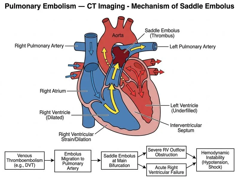
    Sign up free to access AI-powered MCQ practice with detailed explanations and adaptive learning.

    Start Practicing Free More Radiology Questions

    Join our NEET PG community

    Daily MCQs, study tips, and topper strategies on Telegram.

    Join on Telegram →