NEETPGAI
FeaturesNEET PGFMGEINI-CETBlogPricing
Log inStart Free
NEETPGAI

AI-powered NEET PG preparation platform. Master all 19 subjects with adaptive MCQs, AI tutoring, and spaced repetition.

Product

  • Features
  • Subjects
  • Previous Year Questions
  • NEET PG Preparation
  • FMGE Preparation
  • INI-CET Preparation
  • Compare
  • Pricing
  • Blog

Features

  • Adaptive MCQ Practice
  • AI Tutor
  • Mock Tests
  • Spaced Repetition

Resources

  • Blog
  • Study Guides
  • NEET PG Updates
  • Contact & support

Legal

  • Privacy Policy
  • Terms of Service

Stay updated

© 2026 NEETPGAI. All rights reserved.
    Subjects/Anatomy/Radial Nerve — Course and Lesions
    Radial Nerve — Course and Lesions
    medium
    bone Anatomy

    A 32-year-old male labourer presents with inability to extend the thumb at the interphalangeal joint and wrist. On examination, he has wrist drop and sensory loss over the dorsal first web space (between thumb and index finger). He reports a fracture of the mid-shaft humerus 2 weeks ago, which was treated conservatively. Which nerve is injured and at what anatomical level?

    A. Posterior interosseous nerve (PIN) at the level of the supinator muscle
    B. Superficial radial nerve in the distal forearm
    C. Radial nerve at the wrist level within the carpal tunnel
    D. Radial nerve in the spiral groove of the humerus

    Explanation

    Clinical Presentation Analysis

    Key Point
    Mid-shaft humeral fractures are the classic cause of radial nerve injury in the spiral groove, occurring in 10–15% of cases due to the nerve's intimate relationship with the bone in this region.
    Anatomical Correlation

    The radial nerve travels in the spiral groove (radial groove) on the posterior surface of the mid-shaft humerus. A fracture at this level causes direct nerve trauma or compression from callus formation.

    Clinical Features of Radial Nerve Injury at Spiral Groove Level
    Table
    FeatureFindings
    Motor deficitWrist drop (loss of wrist extension via ECRB, ECRL, ECU) + thumb IP extension loss (EPL)
    Sensory lossDorsal first web space (radial-supplied area)
    Nerve branches affectedBoth motor (posterior interosseous nerve) and sensory (superficial radial nerve) branches
    MechanismDirect trauma or compression in spiral groove
    High-YieldNEET PG
    The radial nerve is most vulnerable to injury at the mid-shaft humerus (spiral groove), making humeral fractures the most common cause of radial nerve palsy in clinical practice.
    Why This Level, Not Others?
    Loading diagram...
    Clinical Pearl
    The 2-week timeline and conservative treatment are key: delayed radial nerve palsies can occur from callus compression or nerve entrapment in healing bone.

    Why Correct

    The combination of wrist drop (ECRB/ECRL loss), thumb IP extension loss (EPL loss), and dorsal web space sensory loss (superficial radial nerve) indicates injury to the complete radial nerve at the spiral groove level, where both motor and sensory branches are still together.

    Clinically Oriented Anatomy Moore 8e Ch 6

    Loading illustration…Radial Nerve — Course and Lesions diagram

    Practice similar questions

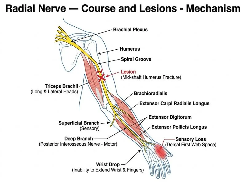
    Sign up free to access AI-powered MCQ practice with detailed explanations and adaptive learning.

    Start Practicing Free More Anatomy Questions

    Join our NEET PG community

    Daily MCQs, study tips, and topper strategies on Telegram.

    Join on Telegram →