NEETPGAI
FeaturesNEET PGFMGEINI-CETBlogPricing
Log inStart Free
NEETPGAI

AI-powered NEET PG preparation platform. Master all 19 subjects with adaptive MCQs, AI tutoring, and spaced repetition.

Product

  • Features
  • Subjects
  • Previous Year Questions
  • NEET PG Preparation
  • FMGE Preparation
  • INI-CET Preparation
  • Compare
  • Pricing
  • Blog

Features

  • Adaptive MCQ Practice
  • AI Tutor
  • Mock Tests
  • Spaced Repetition

Resources

  • Blog
  • Study Guides
  • NEET PG Updates
  • Contact & support

Legal

  • Privacy Policy
  • Terms of Service

Stay updated

© 2026 NEETPGAI. All rights reserved.
    Subjects/Anatomy/Radial Nerve — Course and Lesions
    Radial Nerve — Course and Lesions
    hard
    bone Anatomy

    A 28-year-old man presents with acute-onset severe pain in the lateral aspect of the elbow after a fall on an outstretched hand (FOOSH injury). On examination, he has weakness of finger extension and wrist extension, but normal thumb opposition and sensation in the palm. Sensation is intact over the dorsal first web space. Imaging reveals a fracture-dislocation of the radial head. Which branch of the radial nerve is most likely injured?

    A. Superficial radial nerve (sensory branch)
    B. Deep branch of the radial nerve in the carpal tunnel
    C. Posterior interosseous nerve (PIN) at the level of the supinator muscle
    D. Radial nerve proper in the spiral groove

    Explanation

    Clinical Presentation Analysis

    Key Point
    Radial head fractures and dislocations can entrap or stretch the posterior interosseous nerve (PIN) as it passes through the supinator muscle, causing a pure motor syndrome without sensory loss.
    Anatomical Vulnerability of PIN at Radial Head Level

    The radial nerve divides into its terminal branches distal to the elbow:

    • Superficial radial nerve → sensory (dorsal hand/thumb)
    • Posterior interosseous nerve (PIN) → motor (finger & wrist extension)

    The PIN is particularly vulnerable at the supinator muscle level (just distal to radial head) due to:

    1. 1.
      Passage through the supinator arcade (fibrous tunnel)
    2. 2.
      Proximity to radial head fractures and callus
    3. 3.
      Traction injury from radial head displacement
    Clinical Features of PIN Injury
    Table
    FeaturePIN InjuryRadial Nerve Proper Injury
    Wrist extensionWeak (ECRB, ECRL innervated by PIN)Weak
    Finger extensionWeak/absent (EDC, EIP innervated by PIN)Weak
    Thumb IP extensionWeak (EPL innervated by PIN)Weak
    Thumb oppositionNormal (median nerve)Normal
    Dorsal web space sensationNormal (superficial radial nerve intact)Absent
    Palm sensationNormal (median/ulnar)Normal
    High-YieldNEET PG
    PIN injury produces a pure motor syndrome — no sensory loss because the superficial sensory branch is spared. This is the key discriminator from proximal radial nerve injury.
    Anatomical Course of PIN
    Loading diagram...
    Clinical Pearl
    The supinator muscle acts as a tourniquet around PIN; radial head fractures, swelling, or callus can compress the nerve within this fibrous tunnel, causing delayed or acute PIN palsy ("supinator syndrome").

    Why Correct

    The patient has motor loss (weak finger and wrist extension) but intact sensation (normal dorsal web space sensation and palm sensation). This dissociation—motor loss without sensory loss—is pathognomonic for PIN injury, which is a pure motor nerve. The radial head fracture-dislocation is the anatomical mechanism: PIN passes through the supinator muscle just distal to the radial head and is vulnerable to compression or traction.

    Clinically Oriented Anatomy Moore 8e Ch 6

    Loading illustration…Radial Nerve — Course and Lesions diagram

    Practice similar questions

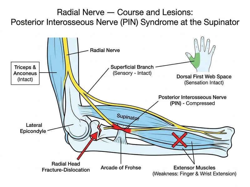
    Sign up free to access AI-powered MCQ practice with detailed explanations and adaptive learning.

    Start Practicing Free More Anatomy Questions

    Join our NEET PG community

    Daily MCQs, study tips, and topper strategies on Telegram.

    Join on Telegram →