NEETPGAI
FeaturesNEET PGFMGEINI-CETBlogPricing
Log inStart Free
NEETPGAI

AI-powered NEET PG preparation platform. Master all 19 subjects with adaptive MCQs, AI tutoring, and spaced repetition.

Product

  • Features
  • Subjects
  • Previous Year Questions
  • NEET PG Preparation
  • FMGE Preparation
  • INI-CET Preparation
  • Compare
  • Pricing
  • Blog

Features

  • Adaptive MCQ Practice
  • AI Tutor
  • Mock Tests
  • Spaced Repetition

Resources

  • Blog
  • Study Guides
  • NEET PG Updates
  • Contact & support

Legal

  • Privacy Policy
  • Terms of Service

Stay updated

© 2026 NEETPGAI. All rights reserved.
    Subjects/Anatomy/Radial Nerve — Course and Lesions
    Radial Nerve — Course and Lesions
    hard
    bone Anatomy

    A 28-year-old woman sustained a stab wound to the proximal forearm just below the elbow. Examination reveals loss of thumb interphalangeal (IP) joint extension and loss of index finger distal interphalangeal (DIP) joint extension, but wrist extension and finger metacarpophalangeal (MCP) joint extension are intact. Which finding best distinguishes this as a posterior interosseous nerve (PIN) injury rather than a more proximal radial nerve lesion at the spiral groove?

    A. Loss of thumb IP extension only
    B. Preserved wrist extension
    C. Intact finger MCP extension
    D. Preserved sensation over the dorsal first web space

    Explanation

    Distinguishing PIN Injury from Proximal Radial Nerve Lesion

    Anatomical Basis

    The radial nerve divides into two branches in the proximal forearm:

    1. 1.
      Superficial radial nerve — sensory to dorsal hand
    2. 2.
      Posterior interosseous nerve (PIN) — motor to finger and thumb extensors

    The wrist extensor muscles (extensor carpi radialis longus and brevis) receive motor innervation from the main radial nerve BEFORE it branches into PIN. This is the critical anatomical distinction.

    Motor Innervation of Extensors
    Table
    MuscleInnervationSpiral Groove LesionPIN Lesion
    ECRL / ECRB (wrist extensors)Radial nerve (proximal)ParalyzedPreserved
    EDC (finger MCP extensors)PINParalyzedParalyzed
    EIP (index DIP extensor)PINParalyzedParalyzed
    EPL (thumb IP extensor)PINParalyzedParalyzed
    EPB (thumb MCP extensor)PINParalyzedParalyzed
    Key Point
    The wrist extensor muscles branch off the radial nerve proximal to the PIN origin. Therefore:
    • Spiral groove lesion → Loss of wrist extension + loss of finger/thumb extension
    • PIN lesion → Preserved wrist extension + loss of finger/thumb extension
    Clinical Presentation in This Case

    The patient has:

    • ✓ Preserved wrist extension ← This is the discriminator
    • ✗ Loss of thumb IP extension (EPL — PIN-innervated)
    • ✗ Loss of index DIP extension (EIP — PIN-innervated)
    • ✓ Intact finger MCP extension (EDC — PIN-innervated, but this is a motor loss, not preserved)
    Clinical Pearl
    The preservation of wrist extension in the presence of finger/thumb extension loss is pathognomonic for a PIN lesion distal to the branching of the wrist extensor nerves. A proximal radial nerve lesion (spiral groove) would cause both wrist and finger extension loss.
    Mnemonic

    "WRIST FIRST" — Wrist extensors branch from radial nerve FIRST (proximal); PIN branches SECOND (distal). PIN injury spares wrist; proximal injury loses both.

    High-YieldNEET PG
    This is a classic NEET PG discriminator question. The single best feature distinguishing PIN from proximal radial nerve injury is preserved wrist extension.

    Loading illustration…Radial Nerve — Course and Lesions diagram

    Practice similar questions

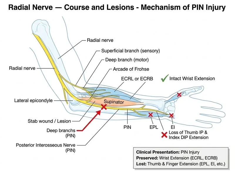
    Sign up free to access AI-powered MCQ practice with detailed explanations and adaptive learning.

    Start Practicing Free More Anatomy Questions

    Join our NEET PG community

    Daily MCQs, study tips, and topper strategies on Telegram.

    Join on Telegram →