## Histopathological Distinction Between ccRCC and pRCC ### Clear Cell RCC (ccRCC) **Key Point:** The hallmark feature of ccRCC is the presence of abundant **intracytoplasmic lipid and glycogen**, which imparts a clear, vacuolated appearance to the cytoplasm on H&E staining. - Cells appear pale or "clear" due to lipid extraction during routine processing - PAS-positive, diastase-resistant glycogen is abundant - Accounts for ~70–80% of all RCC cases - Most aggressive subtype with worst prognosis ### Papillary RCC (pRCC) **Key Point:** pRCC is characterized by a **papillary or tubulopapillary architecture** with cuboidal to columnar epithelial cells, NOT clear cytoplasm. - Cells contain hemosiderin-laden foamy macrophages in the stroma (not in tumor cells) - Low to intermediate nuclear grade typical - Accounts for ~10–15% of RCC cases - Better prognosis than ccRCC ### Comparative Table | Feature | Clear Cell RCC | Papillary RCC | |---------|---|---| | **Cytoplasm** | Clear, lipid/glycogen-rich | Granular, eosinophilic | | **Architecture** | Solid sheets, alveolar | Papillary, tubulopapillary | | **Stromal macrophages** | May be present | Prominent, hemosiderin-laden | | **Frequency** | 70–80% | 10–15% | | **Grade** | Often high (Fuhrman III–IV) | Often low–intermediate | | **Prognosis** | Worse | Better | **High-Yield:** The **clear cell appearance** is the single most reliable discriminator on routine H&E microscopy and is the defining feature that distinguishes ccRCC from all other RCC subtypes, including pRCC. **Clinical Pearl:** ccRCC is associated with VHL gene mutations and can present with paraneoplastic syndromes (erythropoietin, renin, PTHrP secretion), whereas pRCC is often multifocal and bilateral. 
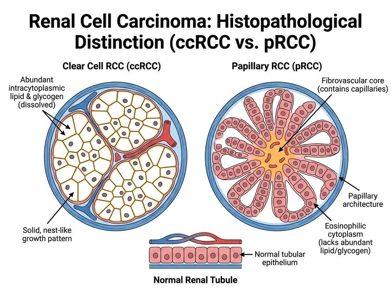
Sign up free to access AI-powered MCQ practice with detailed explanations and adaptive learning.