A 58-year-old Indian man with a 10-year history of chronic kidney disease presents with a left renal mass. Imaging shows a solitary 4 cm mass with minimal fat stranding. On immunohistochemistry, the tumor cells are positive for cytokeratin 7 (CK7) and negative for CD10. Which feature best distinguishes this tumor from clear cell RCC?
A. Positive CK7 and negative CD10 expression pattern
B. Association with chronic kidney disease and acquired cystic kidney disease
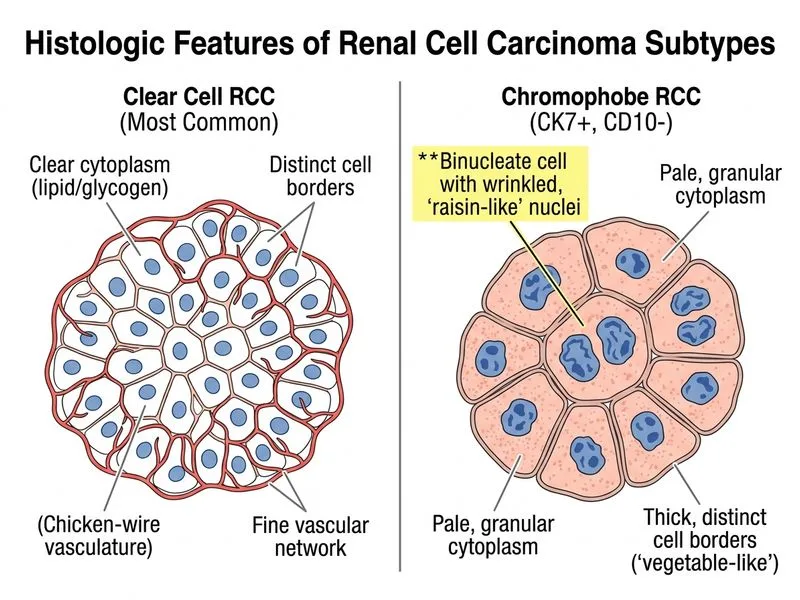
C. Presence of binucleate cells with wrinkled nuclei resembling 'raisin-like' appearance
D. Low Fuhrman nuclear grade with favorable prognosis
Explanation
Chromophobe RCC vs. Clear Cell RCC: Immunohistochemical and Morphological Distinction
Clinical Context
The patient has a solitary renal mass in the setting of chronic kidney disease (CKD). The immunohistochemical profile (CK7+, CD10−) suggests a chromophobe RCC rather than clear cell RCC.
Key Point
While CK7 positivity and CD10 negativity are helpful, the morphological hallmark of chromophobe RCC is the presence of binucleate cells with wrinkled, irregular nuclei resembling "raisin-like" or "vegetable-like" appearance—this is the most reliable discriminator.
Chromophobe RCC Characteristics
Table
Feature
Chromophobe RCC
Clear Cell RCC
Origin
Intercalated cells of collecting duct
Proximal tubular epithelium
Cytoplasm
Pale, granular (flocculent)
Clear, lipid/glycogen-rich
Nuclear morphology
Wrinkled, irregular, "raisin-like"
Round to oval, variable grade
Binucleate cells
Characteristic, frequent
Rare
CK7
Positive
Negative
CD10
Negative
Positive
Hale's colloidal iron
Positive (cell membrane)
Negative
Frequency
5% of RCC
70–80% of RCC
Prognosis
Intermediate (better than ccRCC)
Worst
Association with CKD
Yes (acquired cystic kidney disease)
No
Morphological Discriminator
High-YieldNEET PG
The wrinkled, raisin-like nuclear membrane is the single most distinctive morphological feature of chromophobe RCC on H&E staining. This is pathognomonic and immediately separates it from ccRCC.
Mnemonic
CHROME — Clear cytoplasm (flocculent, not lipid-rich), High binucleate cells, Raisin-like nuclei, Origin from collecting duct intercalated cells, More indolent, Excellent prognosis relative to ccRCC.
Why CK7+/CD10− Is Not the Best Discriminator
Warning
While the immunohistochemical profile (CK7+, CD10−) is useful and supports chromophobe RCC, it is NOT the single best discriminator because:
Some oncocytomas (benign) also show CK7+/CD10−
Immunohistochemistry requires special staining and is not always performed
Morphology on routine H&E is the gold standard and is always available
Clinical Pearl
Chromophobe RCC is associated with Birt-Hogg-Dubé (BHD) syndrome and acquired cystic kidney disease in CKD patients. The patient's 10-year CKD history makes chromophobe RCC more likely than ccRCC.
Loading illustration…
Practice similar questions
Sign up free to access AI-powered MCQ practice with detailed explanations and adaptive learning.