A 32-year-old woman presents with severe pruritus, particularly at night, affecting the interdigital spaces, flexural surfaces, and genitalia. Examination reveals burrows and papules. Which investigation is the gold standard for confirming scabies?
A. Skin biopsy with histopathology
B. Dermoscopy of burrows
C. Scrapings from burrows examined under microscopy for mites, eggs, or feces
D. Fungal culture from lesional material
Explanation
Investigation of Choice for Scabies Confirmation
Key Point
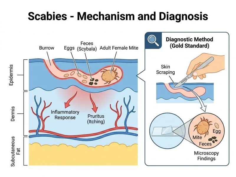
Microscopic examination of scrapings from burrows is the gold standard diagnostic test for scabies, with sensitivity of 60–80% when performed correctly.
Why Scrapings Under Microscopy?
1.
Direct visualization of organisms — Allows identification of the Sarcoptes scabiei mite, eggs, feces (scybala), or eggshells
2.
High specificity — Positive result confirms diagnosis; false positives are rare
3.
Practical and cost-effective — No specialized equipment required; can be done in outpatient clinic
4.
Standard technique:
Select an unscratched burrow (preferably interdigital space, wrist, or genitalia)
Apply mineral oil or potassium hydroxide (KOH) to soften the burrow
Scrape with a blunt blade or needle until slight bleeding occurs
Transfer material to a glass slide
Examine under low power (10× or 40×) for mites, eggs, or feces
Diagnostic Findings on Microscopy
Table
Finding
Appearance
Mite
0.3–0.4 mm, oval, 4 pairs of legs
Eggs
Oval, 100–150 μm
Feces (scybala)
Brown, granular material
Eggshells
Empty, translucent
High-YieldNEET PG
Even a single mite, egg, or fecal pellet on microscopy confirms the diagnosis. Absence does not rule out scabies (sensitivity ~60–80%), but presence is diagnostic.
Clinical Pearl
If microscopy is negative but clinical suspicion remains high (e.g., family members affected, typical distribution), treat empirically — diagnostic certainty should not delay therapy in symptomatic patients.
Sensitivity Considerations
Sensitivity 60–80% — Depends on:
Correct site selection (burrows yield higher sensitivity than papules)
Adequate scraping technique
Number of mites present (lower in treated or partially treated cases)
Examiner experience
Irvine's Dermatology 10e Ch 31
Loading illustration…
Practice similar questions
Sign up free to access AI-powered MCQ practice with detailed explanations and adaptive learning.