## Radiological Pattern of Pulmonary Tuberculosis ### Classic Presentation in Adult TB **Key Point:** Adult-type (reactivation) tuberculosis classically presents with fibrocaseous disease, characterized by cavitation in the apical-posterior segments of the upper lobes. ### Why the Apical-Posterior Segments? 1. **High oxygen tension** — these segments have the highest pO₂, favoring mycobacterial growth 2. **Gravity-dependent pooling** — in upright posture, bacilli settle in these regions 3. **Poor lymphatic drainage** — reduced clearance allows organism proliferation ### Radiological Features of Reactivation TB | Feature | Characteristic | |---------|----------------| | **Location** | Apical-posterior segments of upper lobes (bilateral in ~50%) | | **Pattern** | Fibrocaseous consolidation with cavitation | | **Cavities** | Thin-walled, irregular margins; often multiple | | **Associated findings** | Hilar lymphadenopathy (variable), surrounding infiltrates | | **Pleural involvement** | Minimal unless complicated | ### Clinical-Radiological Correlation - **Sputum smear positivity** + **cavitary disease** = highly infectious - **Upper lobe predominance** + **cavitation** = pathognomonic for reactivation TB - **Night sweats + weight loss** = systemic manifestations of chronic infection **High-Yield:** Cavitary TB in the apical-posterior upper lobe is the hallmark of reactivation tuberculosis in immunocompetent adults. This is the single most tested radiological pattern in chest TB imaging. **Clinical Pearl:** The presence of cavitation indicates active disease with high bacillary load and transmissibility — these patients are infectious and require respiratory isolation. [cite:Harrison 21e Ch 205] 
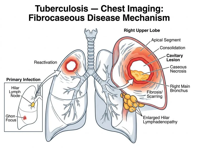
Sign up free to access AI-powered MCQ practice with detailed explanations and adaptive learning.