Ulnar Nerve — Course and Lesions MCQ — NEET PG Practice Question | NEETPGAI
Ulnar Nerve — Course and Lesions
easy
bone Anatomy
At which anatomical location does the ulnar nerve pass through the medial epicondyle of the humerus?
A. Through the carpal tunnel alongside the median nerve
B. Through the pronator teres muscle
C. Between the olecranon process and medial epicondyle
D. Through the cubital tunnel (between the two heads of flexor carpi ulnaris)
Explanation
Ulnar Nerve Course at the Elbow
Anatomical Passage
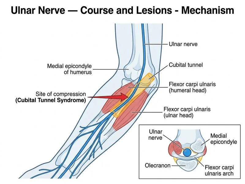
The ulnar nerve passes posterior to the medial epicondyle of the humerus, running through the groove between the olecranon process and the medial epicondyle. This bony groove is the classic "funny bone" location — direct pressure here produces the characteristic tingling sensation along the medial forearm and ring/little fingers.
Key Point
The nerve lies directly in this groove (the epicondylar/ulnar groove), where it is subcutaneous and vulnerable to direct trauma. This is the landmark used clinically to palpate and identify the ulnar nerve at the elbow.
Cubital Tunnel — Distal to the Groove
The cubital tunnel is formed distal to the epicondylar groove, as the nerve passes between the two heads of flexor carpi ulnaris (FCU):
Humeral head of FCU (arising from the medial epicondyle)
Ulnar head of FCU (arising from the olecranon)
The roof is formed by the arcuate ligament (Osborne's ligament) connecting these two heads
The cubital tunnel is the most common site of ulnar nerve entrapment (cubital tunnel syndrome), but the nerve first passes through the epicondylar groove before entering this tunnel.
Why the Other Options Are Wrong
Option A (cubital tunnel between FCU heads): This is the site of cubital tunnel syndrome and is distal to the medial epicondyle groove; the nerve passes through the groove before entering between the FCU heads.
Option B (carpal tunnel with median nerve): The ulnar nerve travels through Guyon's canal at the wrist, entirely separate from the carpal tunnel.
Option D (pronator teres): The median nerve passes between the two heads of pronator teres, not the ulnar nerve.
Clinical Significance
High-YieldNEET PG
The ulnar nerve in the epicondylar groove is vulnerable to:
Direct trauma ("funny bone" injury)
Cubitus valgus deformity (tardy ulnar nerve palsy)
Prolonged pressure (leaning on elbow)
Clinical Pearl
Compression at the groove produces weakness of intrinsic hand muscles (interossei, hypothenar muscles, medial two lumbricals) and sensory loss over the medial 1½ fingers and hypothenar eminence. (Reference: Gray's Anatomy, 41st ed.; Last's Anatomy, 12th ed.)
Loading illustration…
Practice similar questions
Sign up free to access AI-powered MCQ practice with detailed explanations and adaptive learning.