Which histopathological lesion is pathognomonic for acute rheumatic carditis and is characterized by central fibrinoid necrosis surrounded by lymphocytes, plasma cells, and Anitschkow cells?
A. Healed fibrous scar
B. Verrucous vegetation
C. Libman-Sacks endocarditis
D. Aschoff body
Explanation
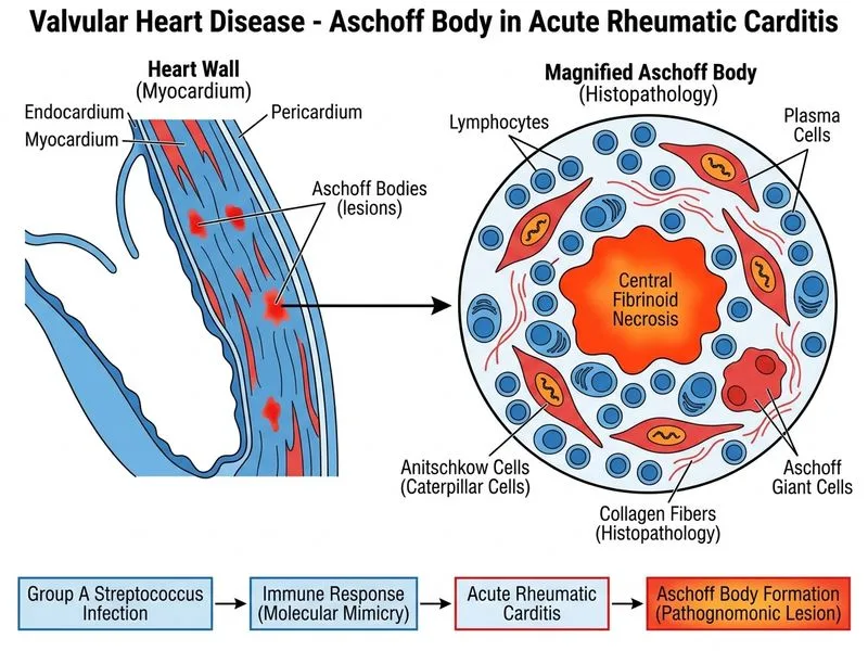
Aschoff Body: The Hallmark of Acute Rheumatic Carditis
Key Point
The Aschoff body is the pathognomonic histological lesion of acute rheumatic carditis. It is a granulomatous lesion found in the myocardium, endocardium, and pericardium.
Microscopic Architecture of an Aschoff Body
Table
Component
Cell Type
Function
Central core
Fibrinoid necrosis
Immune-mediated damage
Inner layer
Anitschkow cells (activated macrophages)
Phagocytosis of necrotic debris
Outer layer
Lymphocytes, plasma cells, fibroblasts
Chronic inflammatory response
Occasional cells
Aschoff giant cells (multinucleated)
Fusion of Anitschkow cells
High-YieldNEET PG
Anitschkow cells are pathognomonic for ARF. They are activated macrophages with a characteristic "caterpillar" or "wavy ribbon" appearance of the nucleus due to wavy chromatin.
Timeline of Aschoff Body Evolution
1.
Acute phase (weeks 1–2): Central fibrinoid necrosis, dense inflammatory infiltrate
Healed phase (months): Replaced by fibrous scar with hyalinization
Clinical Pearl
Aschoff bodies are found in all three layers of the heart (pancarditis), but are most common in the myocardium. Their presence confirms acute rheumatic carditis histologically.
Mnemonic: ASCHOFF — Anitschkow cells + Stratum of lymphocytes + Central fibrinoid necrosis + Hyalinization → Organized fibrous scar (in healing)
Loading illustration…
Practice similar questions
Sign up free to access AI-powered MCQ practice with detailed explanations and adaptive learning.