NEETPGAI
FeaturesNEET PGFMGEINI-CETBlogPricing
Log inStart Free
NEETPGAI

AI-powered NEET PG preparation platform. Master all 19 subjects with adaptive MCQs, AI tutoring, and spaced repetition.

Product

  • Features
  • Subjects
  • Previous Year Questions
  • NEET PG Preparation
  • FMGE Preparation
  • INI-CET Preparation
  • Compare
  • Pricing
  • Blog

Features

  • Adaptive MCQ Practice
  • AI Tutor
  • Mock Tests
  • Spaced Repetition

Resources

  • Blog
  • Study Guides
  • NEET PG Updates
  • Contact & support

Legal

  • Privacy Policy
  • Terms of Service

Stay updated

© 2026 NEETPGAI. All rights reserved.
    Subjects/Pathology/Valvular Heart Disease
    Valvular Heart Disease
    medium
    microscope Pathology

    Which histopathologic feature best distinguishes degenerative (calcific) aortic stenosis from rheumatic aortic stenosis?

    A. Fibrinoid necrosis and inflammatory infiltrate in the valve substance
    B. Calcium deposition confined to the nodules at the base of the cusps with intact commissures
    C. Thickening and retraction of the chordae tendinae
    D. Commissural fusion of valve leaflets

    Explanation

    Practice similar questions

    Sign up free to access AI-powered MCQ practice with detailed explanations and adaptive learning.

    Start Practicing Free More Pathology Questions

    Join our NEET PG community

    Daily MCQs, study tips, and topper strategies on Telegram.
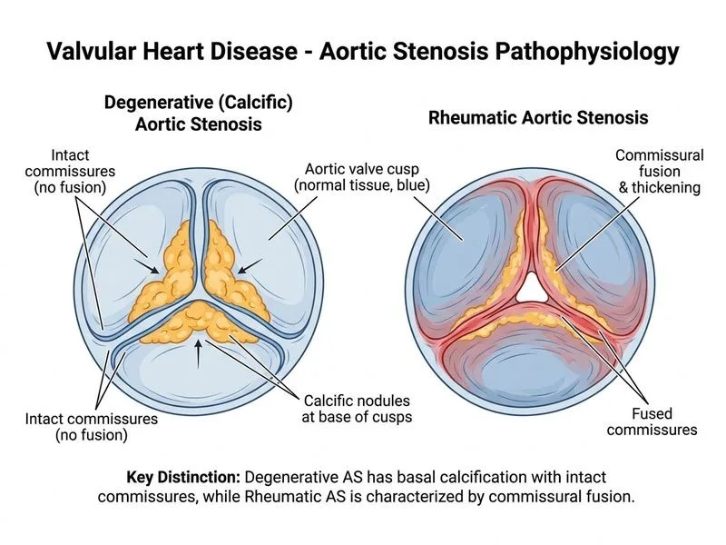
    Join on Telegram →

    Distinguishing Degenerative from Rheumatic Aortic Stenosis

    Pathologic Mechanisms
    Key Point
    Degenerative (calcific) aortic stenosis results from mechanical wear and tear leading to calcium deposition in the aortic cusps, while rheumatic aortic stenosis is an inflammatory sequela of acute rheumatic fever causing commissural fusion and fibrosis.
    Histopathologic Comparison Table
    Table
    FeatureDegenerative (Calcific) ASRheumatic AS
    CommissuresIntact, not fusedFused
    Calcium locationNodules at cusp base (fibrosa side)May be present but not primary
    InflammationMinimal, degenerativeProminent: fibrinoid necrosis, lymphocytes
    Valve substanceFibrosis with calcificationInflammatory infiltrate + fibrosis
    Leaflet mobilityReduced (stiffening)Reduced (retraction + fusion)
    Associated lesionsAortic regurgitation (late)Mitral stenosis (often concurrent)
    Clinical Pearl
    Clinical Pearl
    On gross inspection, degenerative AS shows nodular calcification at the base of the cusps with preserved commissures, whereas rheumatic AS shows commissural fusion with a narrowed orifice. Microscopically, degenerative disease lacks the inflammatory infiltrate characteristic of rheumatic disease.
    High-Yield Discriminator
    High-YieldNEET PG
    Intact commissures with nodular calcification = Degenerative AS. Fused commissures = Rheumatic AS. This distinction is crucial because rheumatic AS is almost always accompanied by mitral valve disease (especially mitral stenosis), whereas degenerative AS is isolated to the aortic valve.
    Mnemonic
    Mnemonic
    DECAL = DEgenerative CALcific (nodules, intact commissures); RHEUM = RHEUMatic (fused commissures, inflammation).

    Robbins 10e Ch 12

    Loading illustration…Valvular Heart Disease diagram